. 2024 Dec 18;10(1):498.
doi: 10.1038/s41420-024-02260-4.

SMURF1 and SMURF2 directly target GLI1 for ubiquitination and proteasome-dependent degradation

Affiliations

SMURF1 and SMURF2 directly target GLI1 for ubiquitination and proteasome-dependent degradation

Fabio Bordin et al. Cell Death Discov. .

Abstract

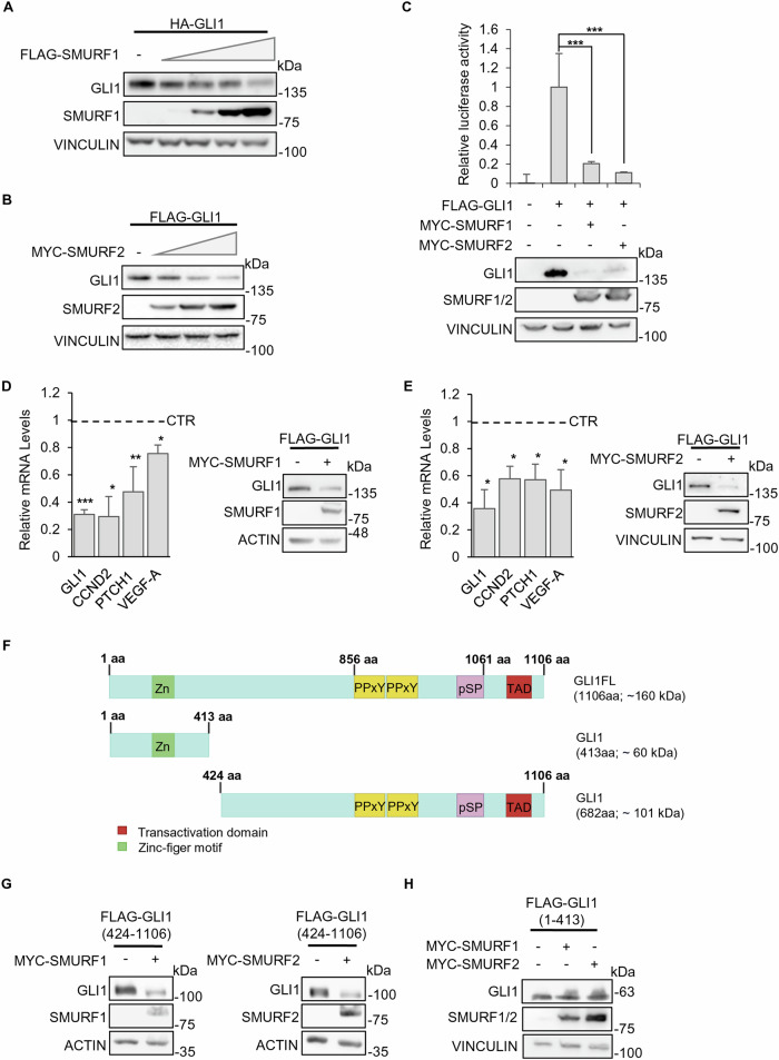
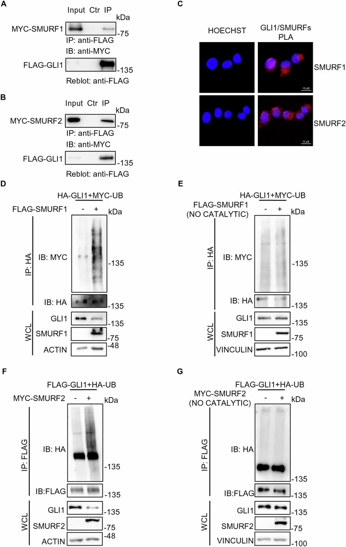
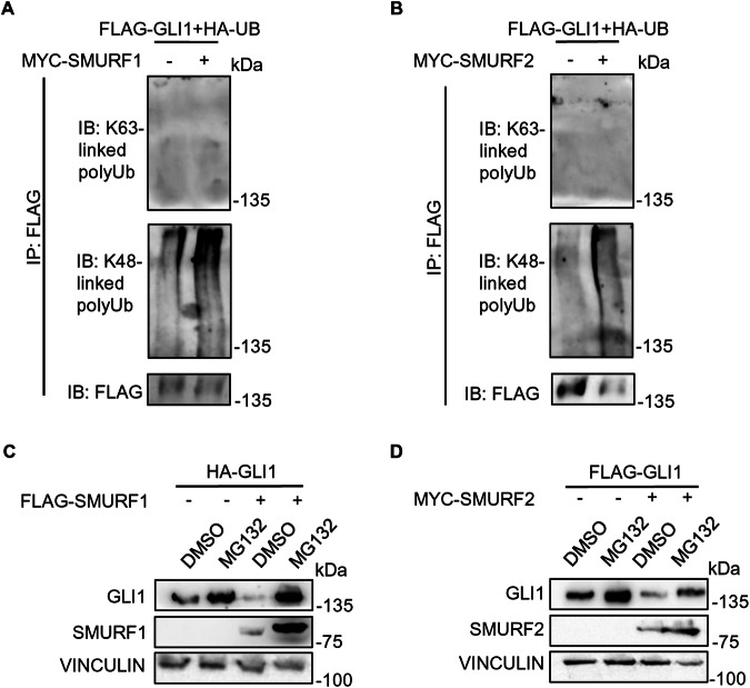
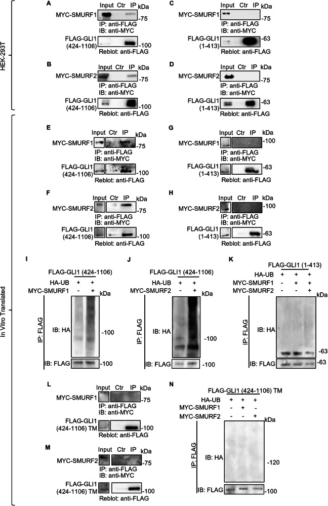
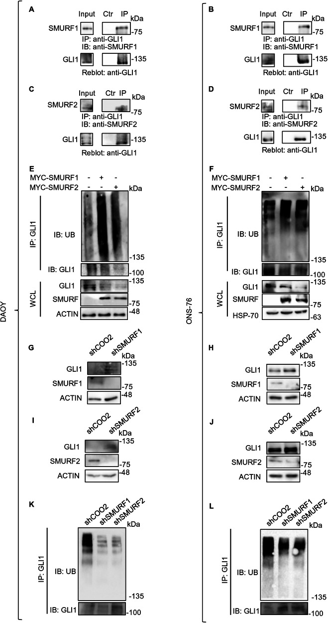
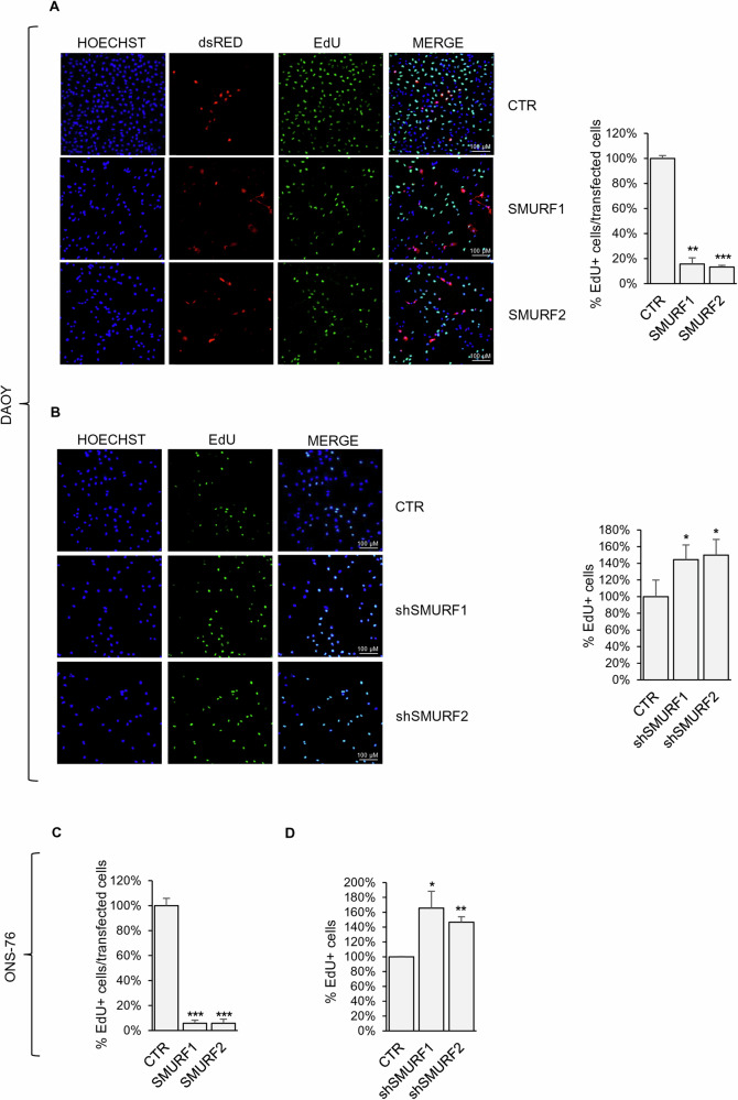
The transcription factor GLI1 is the main and final effector of the Hedgehog signaling pathway, which is involved in embryonic development, cell proliferation and stemness. Whether activated through canonical or non-canonical mechanisms, GLI1 aberrant activity is associated with Hedgehog-dependent cancers, including medulloblastoma, as well as other tumoral contexts. Notwithstanding a growing body of evidence, which have highlighted the potential role of post translational modifications of GLI1, the complex mechanisms modulating GLI1 stability and activity have not been fully elucidated. Here, we present a novel role played by SMURF1 and SMURF2 in the suppression of the Hedgehog/GLI signaling pathway through a direct targeting of GLI1. Indeed, the two SMURFs can interact with GLI1, exploiting the proline rich regions present on GLI1 protein, and trigger its polyubiquitination and proteasomal degradation, leading to a suppression of the Hedgehog pathway activity and a reduction of Hh-dependent tumor cell proliferation. Overall, this study adds new relevance to a tumor suppressive role of SMURFs on the Hedgehog pathway and confers upon them the status of potential therapeutic tools, either in canonical or non-canonical Hedgehog pathway aberrant activation.


Conflict of interest statement

Competing interests: The authors declare no competing interests. Ethics approval and consent to participate: The experiments performed in this work did not use material (i.e., human or mice samples) that requires ethical approval.

Figures

Fig. 1
Fig. 1. SMURFs overexpression reduces GLI1 protein levels.
A, B Effect of SMURFs overexpression on GLI1 protein levels. HEK-293T cells co-expressing HA-GLI1 or FLAG-GLI1 plasmids and increasing amounts of either FLAG-SMURF1, MYC-SMURF2 or control vector. 24 h after transfection the cells were lysed, and proteins analysed using SDS-PAGE. Proteins were detected using antibodies anti-HA, anti-FLAG, anti-MYC and anti-VINCULIN (used as a normalizer). C Luciferase assay using a GLI1-RE activity with or without SMURF1/SMURF2 expression. HEK-293T cells were co-transfected with the vectors Gli1RE-12xLuc and pRL-TK-Renilla (used as a normalizer), along with various combinations of expression vectors FLAG-GLI1, MYC-SMURF1 or MYC-SMURF2 as indicated. 24 h after transfection the cells were lysed for protein expression and luciferase assay. The luciferase results are expressed as a Luciferase/Renilla ratio and were normalized to the control. Protein lysates were analysed using SDS-PAGE, and proteins were detected with antibodies anti-GLI1, anti-MYC and anti-VINCULIN (used as a normalizer). (*p < 0.05, **p < 0.01, ***p < 0.001; results are expressed as the mean ± SD of three independent experiments, Student’s t-test). D, E Analysis on Hh/GLI1 target genes expression, following SMURF1 or SMURF2 expression in HEK-293T cells. 24 h after transfection the cells were lysed for protein and mRNA extraction. mRNA levels of Gli1, Cyclin D2, Ptch1 and VEGF-A were analysed by RT-qPCR and normalized to the average of three housekeeping genes. Protein lysates were analysed by SDS-PAGE, and proteins were detected with antibodies anti-FLAG, anti-MYC, anti-ACTIN and anti-VINCULIN (used as normalizers). (*p < 0.05, **p < 0.01; results are expressed as the mean ± SD of three independent experiments, Student’s t-test). F Schematic representation of structure and domains of full-length(FL) and truncated GLI1 protein. G, H Effect of SMURFs overexpression on protein levels of GLI1 truncated forms. HEK-293T cells co-expressing truncated FLAG-GLI1 (424-1106) or FLAG-GLI1 (1-413), MYC-SMURF1, MYC-SMURF2 or control vector were lysed, and the lysate was analysed using SDS-PAGE. Proteins were detected using antibodies anti-FLAG, anti-MYC, and anti-VINCULIN or anti-ACTIN (used as normalizers).
Fig. 2
Fig. 2. SMURF proteins interact with and increase the ubiquitination levels of full length GLI1 protein.
A, B Co-IP assays on HEK-293T cells expressing FLAG-GLI1 and either MYC-SMURF1 or MYC-SMURF2. 24 h after transfection the cells were lysed, and Co-IP assay was performed using anti-FLAG conjugated agarose beads. As a control, the agarose beads were saturated with the FLAG peptide. Antibodies anti-FLAG and anti-MYC were used to detect the immunocomplexes. C Proximity ligation assay (PLA) for SMURFs and GLI1 interaction. HEK-293T cells were transfected with a construct codifying for FLAG-GLI1. 24 h after transfection, cells were fixed in paraformaldehyde (PFA) and permeabilized. Subsequently, cultures were processed with primary antibody anti-FLAG, anti-GLI1, anti-SMURF1 or anti-SMURF2, and with specific secondary antibodies for PLA assay (red signal), as described in Methods. Nuclei were stained blue (Hoechst). D, G Ubiquitination assays on HEK-293T cells co-expressing HA-GLI1 or FLAG-GLI1, in combination with exogenous Ubiquitin (MYC-UB or HA-UB), and either control vectors, FLAG-SMURF1 (catalytic or not catalytic) or MYC-SMURF2 (catalytic or not catalytic). 24 h after transfection, cells were lysed, and the ubiquitination status was evaluated by immunoprecipitation with anti-HA or anti-FLAG conjugated agarose beads. Proteins were detected using antibodies anti-FLAG, anti-HA, anti-MYC, and anti-ACTIN or anti-VINCULIN (used as a normalizer).
Fig. 3
Fig. 3. SMURF proteins mediate GLI1 K48-poly-ubiquitination, leading to GLI1 proteasomal degradation.
A, B Ubiquitination assays of HEK-293T cells following expression of FLAG-GLI1, HA-UB and either control vectors, MYC-SMURF1 or MYC-SMURF2. 24 h after transfection, cells lysates were immunoprecipitated with FLAG agarose beads and analysed by SDS-PAGE. Proteins were detected using antibodies anti-FLAG, anti-MYC and specific antibodies against K48-linked/K63-linked poly-ubiquitination to detect the ubiquitination status of GLI1. C, D Analysis of GLI1 protein levels following SMURF1/SMURF2 expression in presence of proteasome inhibitor MG132. HEK-293T were transfected with HA-GLI1 or FLAG-GLI1, in combination with control vector, FLAG-SMURF1 or MYC-SMURF2. 24 h post transfection, cells were treated with DMSO or MG132 at 1 µM for 16 h. Subsequently, cells were lysed and analysed by SDS-PAGE. Proteins were detected using antibodies anti-FLAG, anti-HA, anti-MYC, and anti-VINCULIN (used as a normalizer).
Fig. 4
Fig. 4. SMURF proteins interact with the 424-1106 region of GLI1, recognizing its proline rich motifs.
Co-IP assays on HEK-293T cells expressing either MYC-SMURF1 or MYC-SMURF2, together with FLAG-GLI1 (424-1106; A, B) or FLAG-GLI1 (1-413; C, D). 24 h after transfection, cells lysates were used to perform Co-IP assay following immunoprecipitation with anti-FLAG conjugated agarose beads. In the control the agarose beads were saturated with the FLAG peptide. Anti-FLAG and anti-MYC antibodies were used to detect the immunocomplexes. Co-IP assays between in vitro translated (IVT) proteins. MYC-SMURF1 and MYC-SMURF2 together with FLAG-GLI1 (424-1106; E, F) or FLAG-GLI1 (1-413; G, H), were translated as described in Methods and immunoprecipitated using anti-FLAG conjugated agarose beads. In the control, the agarose beads were saturated with the FLAG peptide. Anti-FLAG and anti-MYC antibodies were used to detect the immunocomplexes. In vitro ubiquitination assays using IVT protein. IVT FLAG-GLI1 (424-1106; I, J) or FLAG-GLI1 (1-413; K) were immunoprecipitated by using anti-FLAG conjugated agarose beads and incubated with in vitro ubiquitination components (as indicated in Methods) in the presence of either MYC-SMURF1 or MYC-SMURF2 IVT proteins. Proteins were analysed by SDS-PAGE and detected using antibodies anti-FLAG, anti-MYC, and anti-HA. L, M Co-IP assays between IVT proteins. FLAG-GLI1 (424-1106) triple mutant (TM), MYC-SMURF1 and MYC-SMURF2 IVT proteins were used to perform Co-IP assay following immunoprecipitation with anti-FLAG conjugated agarose beads. In the control, the agarose beads were saturated with the FLAG peptide. Antibodies anti-FLAG and anti-MYC were used to detect the immunocomplexes. N In vitro ubiquitination assays using IVT proteins. FLAG-GLI1 (424-1106) TM IVT was immunoprecipitated by using anti-FLAG conjugated agarose beads and incubated with in vitro ubiquitination components, in the presence or absence of either MYC-SMURF1 or MYC-SMURF2 IVT proteins. Proteins were analysed by SDS-PAGE and detected using antibodies anti-FLAG, anti-MYC, and anti-HA.
Fig. 5
Fig. 5. SMURFs modulation affects GLI1 ubiquitination and protein levels in MB cells.
A, D Endogenous Co-IP assays on DAOY and ONS-76 cells. Cells were lysed and the lysate was immunoprecipitated with either a GLI1 antibody or a control IgG antibody. Antibodies anti-GLI1, anti-SMURF1 and anti-SMURF2 were used to detect the immunocomplexes. E, F Ubiquitination assays on DAOY and ONS-76 cells. Cells were transfected with either control vector, MYC-SMURF1 or MYC-SMURF2. 24 h after transfection, cells were lysed, and the ubiquitination status was evaluated following GLI1 immunoprecipitation. Proteins were detected using antibodies anti-GLI1, anti-SMURF1, anti-SMURF2, anti-Ubiquitin and anti-ACTIN (used as a normalizer) GJ Analysis of GLI1 protein levels following SMURFs silencing. DAOY and ONS-76 cells were stably transduced with the indicated shRNAs against SMURF1 or SMURF2. Cell lysates were analysed using SDS-PAGE. Proteins were detected using antibodies anti-GLI1, anti-SMURF1, anti-SMURF2 and anti-ACTIN (used as a normalizer). K, L Ubiquitination assays on DAOY and ONS-76 cells stably transduced with the indicated shRNAs against SMURF1 or SMURF2. The ubiquitination status was evaluated following GLI1 immunoprecipitation. Proteins were detected using antibodies anti-GLI1 and anti-Ubiquitin.
Fig. 6
Fig. 6. SMURFs expression reduces GLI1 dependent MB cell proliferation.
EdU incorporation staining on MB cells DAOY and ONS-76 expressing SMURF proteins (AC) or stably transduced cells with the indicated shRNAs against SMURF1 or SMURF2 (BD). Percentage of EdU positive cells was calculated over transfected or total cells as indicated. (*p < 0.05; **p < 0.01; ***p < 0.001 results are expressed as the mean ± SD of three independent experiments, Student’s t-test).

References

    1. Zhang Y, Beachy PA. Cellular and molecular mechanisms of Hedgehog signalling. Nat Rev Mol Cell Biol. 2023;24:668–87. - PubMed
    1. Jing J, Wu Z, Wang J, Luo G, Lin H, Fan Y, et al. Hedgehog signaling in tissue homeostasis, cancers, and targeted therapies. Signal Transduct Target Ther. 2023;8:315. - PMC - PubMed
    1. Skoda AM, Simovic D, Karin V, Kardum V, Vranic S, Serman L. The role of the Hedgehog signaling pathway in cancer: a comprehensive review. Bosn J Basic Med Sci. 2018;18:8–20. - PMC - PubMed
    1. Pietrobono S, Gagliardi S, Stecca B. Non-canonical Hedgehog signaling pathway in cancer: activation of GLI transcription factors beyond smoothened. Front Genet. 2019;10:556. - PMC - PubMed
    1. Sigafoos AN, Paradise BD, Fernandez-Zapico ME. Hedgehog/GLI signaling pathway: transduction, regulation, and implications for disease. Cancers. 2021;13:3410. - PMC - PubMed

LinkOut - more resources