. 2024 Dec 18;15(12):908.
doi: 10.1038/s41419-024-07298-x.

Targeting PPARγ via SIAH1/2-mediated ubiquitin-proteasomal degradation as a new therapeutic approach in luminal-type bladder cancer

Affiliations

Targeting PPARγ via SIAH1/2-mediated ubiquitin-proteasomal degradation as a new therapeutic approach in luminal-type bladder cancer

Chih-Chieh Tu et al. Cell Death Dis. .

Abstract

Bladder cancer (BC) is the second most prevalent genitourinary malignancy worldwide. Despite recent approvals of immune checkpoint inhibitors and targeted therapy for muscle invasive or recurrent BC, options remain limited for patients with non-muscle invasive BC (NMIBC) refractory to Bacillus Calmette-Guérin (BCG) and chemotherapy. NMIBC is more frequently classified as a luminal subtype, in which increased PPARγ activity is a key feature in promoting tumor growth and evasion of immunosurveillance. Cinobufotalin is one of the major compound of bufadienolides, the primary active components of toad venom that has been utilized in the clinical treatment of cancer. We herein focused on cinobufotalin, examining its anticancer activity and molecular mechanisms in luminal-type NMIBC. Our results newly reveal that cinobufotalin strongly suppresses the viability and proliferation of luminal BC cells with minimal cytotoxic effects on normal uroepithelial cells, and exhibits significant antitumor activity in a RT112 xenograft BC model. Mechanistically, our sub-G1-phase cell accumulation, Annexin V staining, caspase-3/8/9 activation, and PARP activation analyses show that cinobufotalin induces apoptosis in luminal-type BC cells. Cinobufotalin significantly inhibited the levels of PPARγ and its downstream targets, as well as lipid droplet formation and free fatty acid levels in RT112 cells. PPARγ overexpression rescued RT112 cells from cinobufotalin-induced apoptosis and mitigated the downregulation of FASN and PLIN4. Finally, we show seemingly for the first time that cinobufotalin promotes SIAH1/2-mediated proteasomal degradation of PPARγ in luminal BC cells. Together, these findings compellingly support the idea that cinobufotalin could be developed as a promising therapeutic agent for treating luminal-type NMIBC.


Conflict of interest statement

Competing interests: The authors declare no competing interests. Ethical approval: All animal experiments followed the ethical guidelines, and the protocols have been reviewed and approved by the institutional Animal Care and Use Committee of Taipei Medical University (IACUC approval no: LAC-2021-0471).

Figures

Fig. 1
Fig. 1. In vitro and in vivo anti-tumor activity of cinobufotalin in BC.
BC and normal uroepithelial SV-HUC-1 cells were treated with the indicated concentrations of cinobufotalin for 48 h and cell viability (A) and proliferation (B) determined using the MTT and SRB assays, respectively. Data are expressed as mean ± SD (n = 3) *p < 0.05, **p < 0.01, ***p < 0.001 and ****p < 0.0001 compared with the control group. C, D Athymic nude mice bearing subcutaneously established RT112 xenograft tumors were randomized into four groups (n = 5) and administered the indicated treatments via intraperitoneal injection (i.p.). C Tumor sizes were measured every 2 days during treatment with cinobufotalin or cisplatin. Tumor growth curves are depicted as fold changes compared with tumor volume on day 1 and expressed as means ± SD (n = 5) **p < 0.01 and ****p < 0.0001 compared with vehicle group. D Body weights of mice were measured every 2 days during the drug treatment period. Data are expressed as mean ± SD (n = 5).
Fig. 2
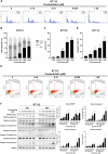
Fig. 2. Cinobufotalin promotes cell cycle accumulation at the sub-G1 phase and apoptosis in RT112 cells.
AC Effects of cinobufotalin on cell cycle distribution in RT112 cells. Cells were treated with different concentrations of cinobufotalin for 48 h and subsequently stained using propidium iodide. The cell cycle was analysed via flow cytometry (A). Quantitative data of histograms (B, C) are expressed as mean ± SD (n = 3) *p < 0.05, ***p < 0.001, and ****p < 0.0001 compared with control group. D, E Effect of cinobufotalin on apoptosis in RT112 cells. Cells were treated with varying concentrations of cinobufotalin for 48 h, stained with Annexin V/7-AAD, and analysed via flow cytometry (D). Quantification of early- and late-stage apoptotic cells (E) are expressed as mean ± SD (n = 3) *p < 0.05, **p < 0.01 and ****p < 0.0001 compared with the control group. F RT112 cells were treated with the indicated concentrations of cinobufotalin for 24 h and 48 h and expression of proteins analysed via western blot. Band intensities of each protein were quantified using Image J and normalized to GAPDH. Fold changes compared to control group are expressed as mean ± SD (n = 3). *p < 0.05, **p < 0.01 and ***p < 0.001 compared with the control group.
Fig. 3
Fig. 3. Effect of cinobufotalin on the transcriptome in RT112 cells.
AC RT112 cells were treated with cinobufotalin (0.625 μM) for 24 h. RNA was extracted using TRIzol reagent, followed by next-generation sequencing (NGS) and bioinformatics analyses (n = 2). A Volcano plot depicts the differentially expressed genes (p adj.<0.05 , |log 2 (Fold Change)| > 1). Red dots indicate upregulation, while blue dots indicate downregulation. The top 10 KEGG pathways displaying the most significant enrichment in differentially expressed genes (B), and the PPAR signaling pathway is significantly enriched (C). D Effects of cinobufotalin on mRNA levels of PPARγ and its downstream target genes in RT112 cells. Cells were treated with the indicated concentrations of cinobufotalin for 24 h and mRNA expression determined via RT-qPCR. Data are expressed as mean ± SD (n = 2). *p < 0.05, **p < 0.01, ***p < 0.001 and ****p < 0.0001 compared with control. E Western blot analysis of protein expression of PPARγ and its downstream targets in different BC and normal uroepithelial SV-HUC-1 cells. F Band intensities of each protein, determined using Image J software. After normalization to the intensity of HSP90, relative protein levels were determined compared with the SV-HUC-1 group. G RT-qPCR analysis of relative mRNA expression levels of PPARγ and its downstream targets in different cell lines compared to normal uroepithelial SV-HUC-1 cells. Data are expressed as mean ± SD (n = 3). *p < 0.05, **p < 0.01, ***p < 0.001 and ****p < 0.0001 compared with SV-HUC-1 cells.
Fig. 4
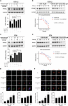
Fig. 4. PPARγ downregulation contributes to cinobufotalin-induced apoptosis in luminal BC cells.
Cinobufotalin downregulates the protein levels of PPARγ and its downstream targets in luminal BC cells. RT112 (A) and RT4 (B) cells were treated with different concentrations of cinobufotalin (0.15–1.25 μM) for 24 h and 48 h or 0.625 μM cinobufotalin over a range of time-points (30 min to 48 h) and subjected to western blot analysis. C Effect of PPARγ overexpression on cinobufotalin-mediated cytotoxicity in RT112 cells. Cells were transiently transfected with pcDNA 3.1 or PPARγ-Flag plasmids and treated with the indicated concentrations of cinobufotalin for 48 h. Cell viability was determined using the MTT assay. Data are expressed as mean ± SD (n = 4) #p < 0.05, ##p < 0.01, and ###p < 0.001 PPARγ-overexpressing group compared with pcDNA 3.1 group. D Overexpression of PPARγ in RT112 cells reverses cinobufotalin-induced apoptosis. Cells were transiently transfected with pcDNA 3.1 or PPARγ-Flag plasmid and treated with the indicated concentrations of cinobufotalin for 48 h. Protein expression was analysed via western blot. E Effect of PPARγ knockdown on the proliferation rate of RT112 cells. PPARγ was stably depleted in RT112 cells with two different sequences of shRNA (673, 926), and cell proliferation rates determined using the MTT assay. Data are expressed as mean ± SD (n = 4). *p < 0.05, **p < 0.01 and ***p < 0.001 compared with the 0-h group ; #p < 0.05, ##p < 0.01 and ###p < 0.001 compared with WT group. Knockdown of PPARγ in RT112 (F) and RT4 (G) cells potentiates cinobufotalin-induced apoptosis. Stable knockdown of PPARγ was achieved with two different sequences of shRNA (673, 926), followed by treatment with cinobufotalin for 48 h. Protein expression was analysed via western blot. Data from statistical analysis of western blots are presented in Supplementary Fig. S7.
Fig. 5
Fig. 5. Cinobufotalin promotes the proteasomal degradation of PPARγ in luminal BC cells.
AD The 20S proteosome inhibitor bortezomib reverses the downregulation of PPARγ induced by cinobufotalin in BC cells. RT112 (A) and RT4 (C) cells were pretreated with bortezomib (0.1 and 1 μM) for 1 h, followed by cinobufotalin (0.625 μM) for 3 h. Protein levels of PPARγ were analysed via western blot and the band intensities of each protein evaluated using Image J software. Data are expressed as mean ± SD (n = 3) ***p < 0.01 and ****p < 0.0001 compared with control; #p < 0.05, ##p < 0.01 compared with cinobufotalin alone (0.625 μM). Cinobufotalin decreased the half-life of PPARγ in BC cells. RT112 (B) and RT4 (D) cells were treated with cycloheximide (40 μM) in the presence or absence of cinobufotalin (0.625 μM) for the indicated times (0.5 h–4 h). Proteins levels were analysed via western blot and the band intensities of each protein determined using Image J software. Data are expressed as mean ± SD (n = 3) *p < 0.05, **p < 0.01 and ***p < 0.001 compared with cycloheximide alone. Cinobufotalin promotes ubiquitin-proteasomal degradation of PPARγ in luminal BC cells. RT112 (E) and RT4 (F) cells were treated with cinobufotalin (0.625 μM) for the indicated times and subjected to immunofluorescence staining and confocal microscopy. Data are expressed as mean ± SD (n = 3) *p < 0.05, **p < 0.01, ***p < 0.001, ****p < 0.0001 compared with 0 h group.
Fig. 6
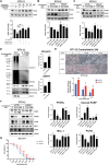
Fig. 6. Identification of the E3 ubiquitin ligase mediating cinobufotalin-induced proteasomal degradation of PPARγ in luminal BC cells.
A The SIAH1/2 inhibitor reverses cinobufotalin-induced degradation of PPARγ in RT112 cells. Cells were pre-incubated with menadione for 30 min, followed by treatment with cinobufotalin for 3 h. Western blot analysis was conducted and the band intensities of each protein determined using Image J software. Data are expressed as mean ± SD (n = 3) *p < 0.05 and ****p < 0.0001 compared with control, ##p < 0.01 compared with cinobufotalin 0.625 μM alone. BD SIAH1 significantly contributes to the cinobufotalin-induced ubiquitin-proteasomal degradation of PPARγ in RT112 cells. RT112 cells were transfected with non-targeting control (siCTRL), siRNA targeting SIAH1 (B) or SIAH2 (C), and exposed to the indicated concentrations of cinobufotalin for 3 h. Cells were subjected to western blot analysis and the band intensities of each protein determined using Image J software. Data are expressed as mean ± SD (n = 3) **p < 0.01 and ***p < 0.001, compared with control, #p < 0.05 and ##p < 0.01 compared with the siCTRL group. D RT112 cells were exposed to cinobufotalin (0.625 μM) for 2 h and subjected to immunoprecipitation using an antibody against PPARγ, followed by western blot analysis. Band intensities of each protein were determined using Image J software. Data are expressed as mean ± SD (n = 3) ***p < 0.001 compared with control (CTRL). E, F SIAH1 is crucial to cinobufotalin-induced lipid droplet suppression and apoptosis. RT112 cells were transfected with non-targeting control (siCTRL) or siRNA targeting SIAH1 (siSIAH1), and treated with cinobufotalin for 24 h and subjected to Oil Red O staining (E). The relative Oil red O-positive areas were analysed with the Image J software. Data are expressed as mean ± SD (n = 3) ***p < 0.001 compared with control, #p < 0.05, ##p < 0.01 compared with the siCTRL group. F RT112 cells were transiently transfected with non-targeting control (siCTRL) or siSIAH1. After transfection, the cells were exposed to cinobufotalin for 48 h, and subjected to Western blot analysis. Band intensities of each protein were determined by using the Image J software and normalized to HSP90. Data are expressed as mean ± SD (n = 3) *p < 0.05, **p < 0.01, ***p < 0.001 and ****p < 0.0001 compared with control group; #p < 0.05, ##p < 0.01 and ###p < 0.001 compared with siCTRL group. G The effect of SIAH1 knockdown on cinobufotalin-mediated cytotoxicity in RT112 cells. The cells were transiently transfected with control (siCTRL) or SIAH1 siRNA, and treated with indicated concentrations of cinobufotalin for 48 h. Cell viability was assessed using MTT assay. Data are expressed as mean ± SD (n = 3) *p < 0.05, **p < 0.01 and ****p < 0.0001 compared to control group; ##p < 0.01 and ###p < 0.001 compared to the siCTRL group.
Fig. 7
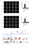
Fig. 7. Cytosolic distribution of SIAH1 is crucial to cinobufotalin-induced ubiquitination of PPARγ in RT112 cells.
RT112 cells were treated with cinobufotalin (0.625 μM) in the presence or absence of menadione (40 μM) for 2 h and subjected to immunofluorescence staining with antibodies against ubiquitin (Ub) and PPARγ (A) or SIAH1 and SIAH2 (B). The images were acquired by confocal microscopy, and the data are expressed as mean ± SD (n = 3) ***p < 0.001 compared with CRTL group; ###p < 0.001 compared with cino 0.625 μM group. C Proposed anticancer mechanism of cinobufotalin in luminal-type BC cells. Cinobufotalin induces apoptosis by promoting the ubiquitin-proteasomal degradation of PPARγ, facilitated by E3 ligases SIAH1 and SIAH2 in the cytoplasm and nucleus, respectively. The decrease in PPARγ levels leads to the suppression of its downstream targets, FASN and PLIN4, which in turn reduces fatty acid synthesis and lipid droplet formation, potentially activating caspase-dependent apoptosis in luminal BC cells. Furthermore, cinobufotalin exhibits significant antitumor activity in a RT112 xenograft BC model in vivo. PPRE: PPAR response element. The schematic representation was generated using Biorender (©BioRender-biorender.com, San Francisco, CA, USA).

References

    1. Sung H, Ferlay J, Siegel RL, Laversanne M, Soerjomataram I, Jemal A, et al. Global cancer statistics 2020: GLOBOCAN estimates of incidence and mortality worldwide for 36 cancers in 185 countries. CA Cancer J Clin. 2021;71:209–49. - PubMed
    1. Babjuk M, Burger M, Zigeuner R, Shariat SF, van Rhijn BW, Comperat E. et al. EAU guidelines on non-muscle-invasive urothelial carcinoma of the bladder: update 2013. Eur Urol. 2013;64:639–53. 10.1016/j.eururo.2013.06.003. - PubMed
    1. Joyce DD, Sharma V, Williams SB. Cost-effectiveness and economic impact of bladder cancer management: an updated review of the literature. Pharmacoeconomics. 2023;41:751–69. 10.1007/s40273-023-01273-8. - PubMed
    1. Hannouneh ZA, Hijazi A, Alsaleem AA, Hami S, Kheyrbek N, Tanous F. et al. Novel immunotherapeutic options for BCG-unresponsive high-risk non-muscle-invasive bladder cancer. Cancer Med. 2023;12:21944–68. 10.1002/cam4.6768. - PMC - PubMed
    1. Balasubramanian A, Gunjur A, Weickhardt A, Papa N, Bolton D, Lawrentschuk N. et al. Adjuvant therapies for non-muscle-invasive bladder cancer: advances during BCG shortage. World J Urol. 2022;40:1111–24. 10.1007/s00345-021-03908-x. - PubMed