. 2024 Dec 18;15(1):10684.
doi: 10.1038/s41467-024-54913-8.

Design of pseudosymmetric protein hetero-oligomers

Affiliations

Design of pseudosymmetric protein hetero-oligomers

Ryan D Kibler et al. Nat Commun. .

Abstract

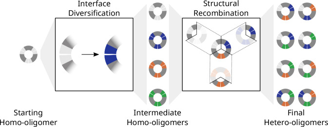
Pseudosymmetric hetero-oligomers with three or more unique subunits with overall structural (but not sequence) symmetry play key roles in biology, and systematic approaches for generating such proteins de novo would provide new routes to controlling cell signaling and designing complex protein materials. However, the de novo design of protein hetero-oligomers with three or more distinct chains with nearly identical structures is a challenging unsolved problem because it requires the accurate design of multiple protein-protein interfaces simultaneously. Here, we describe a divide-and-conquer approach that breaks the multiple-interface design challenge into a set of more tractable symmetric single-interface redesign tasks, followed by structural recombination of the validated homo-oligomers into pseudosymmetric hetero-oligomers. Starting from de novo designed circular homo-oligomers composed of 9 or 24 tandemly repeated units, we redesigned the inter-subunit interfaces to generate 19 new homo-oligomers and structurally recombined them to make 24 new hetero-oligomers, including ABC heterotrimers, A2B2 heterotetramers, and A3B3 and A2B2C2 heterohexamers which assemble with high structural specificity. The symmetric homo-oligomers and pseudosymmetric hetero-oligomers generated for each system have identical or nearly identical backbones, and hence are ideal building blocks for generating and functionalizing larger symmetric and pseudosymmetric assemblies.


Conflict of interest statement

Competing interests: A provisional patent application has been filed (63/486,872) by the University of Washington, listing R.D.K., B.I.M.W., B.L.S., S.L., and D.B. as inventors or contributors. The remaining authors declare no competing interests.

Figures

Fig. 1
Fig. 1. Overview of pseudosymmetric structural recombination design approach.
Diagram of our stepwise hetero-oligomer creation strategy. Flowing left to right, the interface of the starting parental homo-oligomer is redesigned to generate variant homo-oligomeric interfaces, followed by junction splicing to assemble them into hetero-oligomers. Cartoons represent trimers with colored interfaces that match up (separated by a small white gap in the cartoon) and have unique sequences according to their color. Interfaces on the same chain are connected via a homology region in dark gray, which has the same sequence and structure across all protomer variants. In this strategy, the interface of an existing homo-oligomer (left; a homotrimer in this example) is redesigned symmetrically (termed Interface Diversification), with the junction regions remaining unchanged. Homo-oligomers that assemble correctly (middle column) are cut at the same point within their common homology regions and then spliced back together (termed Structural Recombination), making two-chain hybrids with two different interfaces (Supplementary Fig. 3b) which assemble into heterotrimers (right column). It is possible to create two different heterotrimers with the same interfaces by rearranging the order of the interfaces (Supplementary Fig. 3c).
Fig. 2
Fig. 2. Characterization of BGL homotrimers.
a Cartoon representation of six representative successful redesigns of BGL0, with two from each design set: Helix Rebuilding plus C-terminal attachment (HR-C), Helix Rebuilding plus N-terminal attachment (HR-N), and Normal Modes (NM) relaxation. Blue and orange colored backbones highlight N- and C-terminal two-helix segments comprising the interface, which was redesigned, and newly designed hydrogen bond networks (HBNets) are shown in sticks with the hydrogen bonds in dashed lines. Design names in the bottom left corner of each box and box outline and trace colors are used consistently throughout. Characterization of the six examples from (a) by (b) SEC, (c) SAXS, (d) nsEM, and (e) nMS. b Overlay of SEC traces normalized to maximum absorbance at 280 nm (A280), after soluble aggregate removal and TEV cleavage. SEC traces of all 20 designs are provided in Supplementary Fig. 4. c Overlay of SAXS data. Data were scaled appropriately to align at q = 0.01 to highlight the similarity of the profiles. d Individual 2D class averages of nsEM data for each sample. e Individual mass deconvolved nMS data of TEV-cleaved samples (Supplementary Data 13). All peaks that match the expected mass in Daltons for each homotrimer are annotated with green circles, and homohexamers (likely to be dimers of trimers) are annotated with orange diamonds.
Fig. 3
Fig. 3. Crystal structures of BGL homotrimers.
a, b, d Crystal structures (white) of four different BGL homotrimers are superimposed onto the design models colored in teal, purple, and magenta. (left) The top-down view of the full ring with HBNets highlighted and visible through transparent cartoon models. The backbones of all crystal structures match well with the design models (<1.4 Å RMSD). (Right) Close-up view(s) of each HBNet well enough resolved to build full side chains. a BGL06 (2.1 Å resolution; RMSD = 1.2 Å over all 483 Cα atoms; PDB: 8E0L) and (d) BGL18 (3.0 Å resolution; RMSD = 1.4 Å over all 513 Cα atoms; PDB: 8E0N) were both well enough resolved to model all three interfaces. BGL18 contained two copies of the full trimer in the crystal unit cell, which are structurally similar to each other (RMSD = 0.3 Å over all 511 Cα atoms between copies). b One interface of BGL14 (3.0 Å resolution; RMSD = 1.3 Å over all 487 Cα atoms; PDB: 8E12) was well resolved. c BGL15 (4.0 Å resolution; PDB: 8E0M) had three copies of the trimer in the unit cell and all three were close to the design model (0.6 Å (519 residues), 0.6 Å (519 residues), and 0.7 Å (519 residues) RMSD over Cα atoms).
Fig. 4
Fig. 4. Characterization of hetBGL heterotrimers.
ae Results of hetBGL03-15-18. a (top) Schematic showing the result of structural recombination between BGL03, BGL15, and BGL18. (bottom) The expression constructs for each chain as expressed tricistronically. GFP is superfolderGFP, included to add a large amount of mass and provide an additional spectroscopic identification, and MP is EHEE_rd2_0005, a stable and small protein included to add a small amount of mass. b SDS-PAGE of IMAC elution. M: protein ladder. E: IMAC elution. Staining intensity follows the trend expected for three proteins of very different masses at equal abundance (larger proteins binding more stain). Representative image showing similar results found from three separate purifications. Source data are provided as a Source Data file. c SEC trace overlaying normalized absorbances at A280 and A485. d Mass deconvolved nMS data showing all annotated species (Supplementary Data 13). The insert plot is the zoomed-in region indicated by the dashed box on the main plot, showing a 24 kDa wide region centered on the ABC peak on the x-axis and with signal intensity between 0% and 10% on the y-axis. Shaded regions in red indicate the expected locations for off-target trimers. e (left) Crystal structure in white (2.1 Å resolution, PDB: 8E0O) and design model in teal, purple, and magenta overlaid to show global agreement (Cα-RMSD: 1.2 Å over 506 residues, TM-score: 0.96). Dashed boxes indicate approximate locations of zoomed-in views of the interfaces derived from BGL03, BGL15, and BGL18, showing side chains of HBNet residues and their inferred hydrogen bonds as yellow dashes. f Individual mass deconvolved nMS data for all hetBGL Set 2 samples (Supplementary Data 13). Peaks labeled unk are unknown species believed to be distinct from noise.
Fig. 5
Fig. 5. Characterization of RTR homotetramers and hetero-oligomers.
a Cartoon representation of three representative successful redesigns of RTR0. Blue and purple colored backbones represent N- and C-terminal interfaces, respectively. HBNets and disulfide bonds are shown in sticks with hydrogen bonds in dashed lines. Design names (bottom left corner of each box) and box outline and trace colors are used consistently throughout to represent the same interfaces. Characterization of the three examples from (a) by (b) SEC, c nMS, d SAXS, and (e) nsEM. b Overlay of A280 normalized SEC traces. c Cropped view from one of 173 micrographs of RTR05 collected from a single sample. d Individual 2D class averages of nsEM data collected on each sample. The outline color corresponds to the sample ID. e SEC traces (normalized absorbance at 230 nm) of four hetRTRs: (left to right) A2B2-hetRTR05-11noCys, A52B72-hetRTR05-11noCys, A3B3-hetRTR05-11noCys, and A2B2C2-hetRTR11noCys-05-16. Inset with cartoons representing the expected oligomer shapes. Shading indicates the region used for nsEM analysis. f Pairs of 2D class averages matching the SEC traces above, except A52B72-hetRTR05-11noCys which shows one 2D class average and an overlay of the 2D class with a 2D class of RTR11 to show the angular shift.

Update of

References

    1. Levy, E. D. & Teichmann, S. A. Chapter two - structural, evolutionary, and assembly principles of protein oligomerization. In Oligomerization in Health and Disease (eds. Giraldo, J. & Ciruela, F.) 25–51 (Academic Press, 2013). - PubMed
    1. Lang, D., Thoma, R., Henn-Sax, M., Sterner, R. & Wilmanns, M. Structural evidence for evolution of the β/α barrel scaffold by gene duplication and fusion. Science289, 1546–1550 (2000). - PubMed
    1. Wilusz, C. J. & Wilusz, J. Lsm proteins and Hfq: life at the 3’ end. RNA Biol.10, 592–601 (2013). - PMC - PubMed
    1. Khusial, P., Plaag, R. & Zieve, G. W. LSm proteins form heptameric rings that bind to RNA via repeating motifs. Trends Biochem. Sci.30, 522–528 (2005). - PubMed
    1. Fromont-Racine, M. et al. Genome-wide protein interaction screens reveal functional networks involving Sm-like proteins. Yeast17, 95–110 (2000). - PMC - PubMed

Publication types

LinkOut - more resources