. 2025 Jul;643(8070):192-200.
doi: 10.1038/s41586-024-08329-5. Epub 2024 Dec 18.

Lithocholic acid phenocopies anti-ageing effects of calorie restriction

Affiliations

Lithocholic acid phenocopies anti-ageing effects of calorie restriction

Qi Qu et al. Nature. 2025 Jul.

Erratum in

Abstract

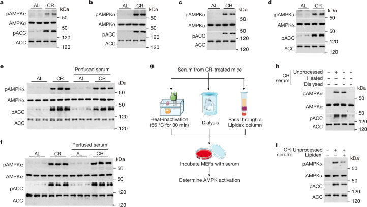
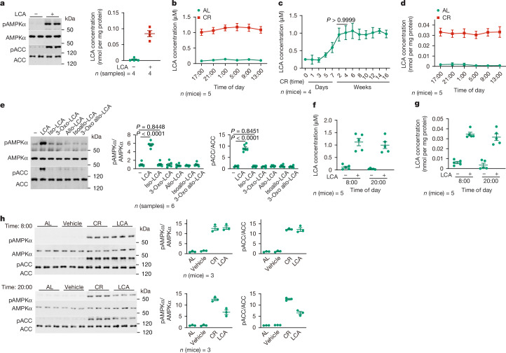
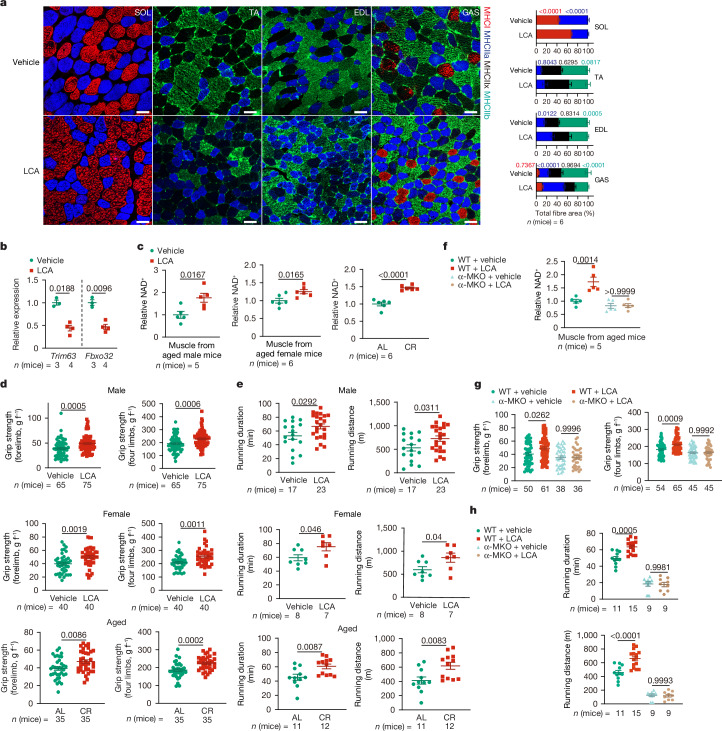
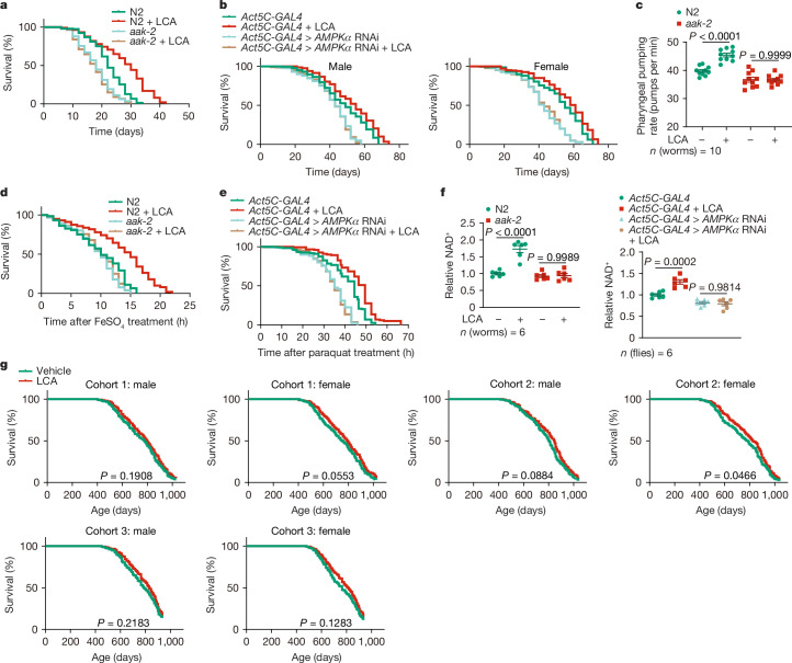
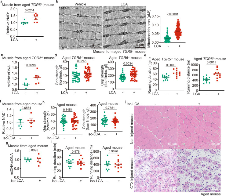
Calorie restriction (CR) is a dietary intervention used to promote health and longevity1,2. CR causes various metabolic changes in both the production and the circulation of metabolites1; however, it remains unclear which altered metabolites account for the physiological benefits of CR. Here we use metabolomics to analyse metabolites that exhibit changes in abundance during CR and perform subsequent functional validation. We show that lithocholic acid (LCA) is one of the metabolites that alone can recapitulate the effects of CR in mice. These effects include activation of AMP-activated protein kinase (AMPK), enhancement of muscle regeneration and rejuvenation of grip strength and running capacity. LCA also activates AMPK and induces life-extending and health-extending effects in Caenorhabditis elegans and Drosophila melanogaster. As C. elegans and D. melanogaster are not able to synthesize LCA, these results indicate that these animals are able to transmit the signalling effects of LCA once administered. Knockout of AMPK abrogates LCA-induced phenotypes in all the three animal models. Together, we identify that administration of the CR-mediated upregulated metabolite LCA alone can confer anti-ageing benefits to metazoans in an AMPK-dependent manner.


Conflict of interest statement

Competing interests: The authors declare no competing interests.

Figures

Fig. 1
Fig. 1. Serum from CR-treated mice can activate AMPK in cells and in mice.
ad, Serum from CR-treated mice can activate AMPK in cells cultured in normal medium. MEFs (a), HEK293T cells (b), primary hepatocytes (c) and primary myocytes (d) were cultured in DMEM (a,b), William’s medium E (c) or Ham’s F-10 medium (d). However, FBS (a,b,d) or BSA (c) supplemented in the culture medium was replaced with an equal volume of serum from mice subjected to CR for 4 months (collected at 17:00, immediately before the next food supply; this CR serum was used hereafter, unless stated otherwise) or serum from mice on an ad libitum diet, for 4 h (AL serum; control). Cells were then lysed, followed by determination of AMPK activation by immunoblotting (IB) for pAMPKα and pACC levels. e,f, Serum from CR-treated mice activates AMPK in the muscle and liver of mice perfused with this serum. Ad libitum-fed mice were perfused through the jugular with 100 μl of CR serum or AL serum (as control), followed by determination of AMPK activation in liver (e) and muscle tissues (f) at 2 h after perfusion by IB for pAMPKα and pACC. gi, Heat-stable, polar metabolites with low molecular weights in CR serum mediate AMPK activation. Schematic of experiment (g). MEFs were treated with CR serum as in a, except that the serum was heat-inactivated, dialysed or passed through a Lipidex column, followed by determination of AMPK activation by IB (h,i). Experiments were performed three (c–i) or five (a,b) times. Artwork in g was reproduced from Servier Medical Art under a Creative Commons Attribution 3.0 unported licence.
Fig. 2
Fig. 2. LCA is increased after CR and responsible for AMPK activation.
a, LCA is the AMPK-activating factor in CR serum. MEFs were treated with 1 μM LCA, a concentration roughly equivalent to that in the serum of CR mice, for 4 h, followed by determination of AMPK activity (left) and intracellular concentrations of LCA (right). bd, Metabolomics analyses reveals substantial increases of LCA in CR-treated mice. Serum (b,c) and muscular (d) concentrations of LCA in mice subjected to CR for 4 months were determined at different time points of the day (b,d) or at different time durations of CR (c). e, Immunoblot (left) and quantification (right) showing that LCA is the sole derivative of bile acids that can activate AMPK. MEFs were treated with 1 μM LCA or 1 μM LCA derivatives for 4 h, followed by determination of AMPK activities. f,g, Mice treated with LCA through drinking water have similar levels of LCA to that in CR serum. Aged, ad libitum-fed (1.5-year-old) mice were fed (2-hydroxypropyl)-β-cyclodextrin-coated LCA at 1 g l–1 in drinking water for 1 month, and concentrations of LCA in serum (f) and muscle tissue (g) of mice at two different times of the day (8:00, representing the light cycle, and 20:00, representing the dark cycle), were measured. h, Immunoblots (left) and quantification (right) showing that LCA administration activates AMPK in mice. Aged mice were subjected to CR as in b, or treated with LCA as in f, followed by determination of AMPK activities in skeletal muscle at both light and dark cycles. Statistical results are shown as the mean ± s.e.m. Specific numbers of mice or samples used are labelled on each panel. P values were calculated using two-way analysis of variance (ANOVA) followed by Tukey’s test (c) or two-sided Student’s t-test (e). Experiments were performed three (be) or four (a,f,g) times. Source Data
Fig. 3
Fig. 3. LCA exerts rejuvenating effects that depend on AMPK.
a,b, LCA induces oxidative fibre conversion and prevents muscle atrophy in aged mice. Ad libitum-fed mice were given (2-hydroxypropyl)-β-cyclodextrin-coated LCA at 1 g l–1 in drinking water for 1 month, followed by determination of muscle fibre type by immunohistochemistry (a) and mRNA levels of the atrophy markers Trim63 and Fbxo32 by RT–PCR (b). EDL, extensor digitorum longus; GAS, gastrocnemius; SOL, soleus; TA, tibialis anterior. Scale bars, 50 µm. c, LCA increases NAD+ levels in aged mice to a level similar to that induced by CR. Ad libitum-fed male (left) and female (middle) mice were either treated with LCA as in a or subjected to CR for 3.5 months (right), followed by determination of muscular NAD+ levels. d,e, LCA promotes muscle strength and endurance in aged mice to an extent similar to that induced by CR. Ad libitum-fed mice, both male and female, were treated with LCA as in a or subjected to CR for 3.5 months, followed by determination of grip strength (d) and running duration (e). f, AMPK is required for the increase in muscular NAD+ by LCA. Ad libitum-fed, aged mice with AMPKα specifically knocked out in muscle (α-MKO) and wild-type (WT) littermates were treated as in a, followed by determination of muscular NAD+ levels. g,h, Muscle-specific AMPK knockout abolishes the effects of LCA on muscle strength and endurance. Mice were treated as in a, followed by determination of grip strength (g) and running duration (h) as in d and e, respectively. Statistical results are shown as the mean ± s.e.m. Specific numbers of mice used are labelled on each panel. P values (shown on the charts) were calculated using two-way ANOVA followed by Tukey’s test (a,fh) or two-sided Student’s t-test (be). Experiments were performed three times. Source Data
Fig. 4
Fig. 4. LCA extends lifespan and healthspan.
a,b, LCA extends lifespan in nematodes and flies through AMPK. WT (N2) or aak-2 knockout nematodes, (a) and WT (Act5C-GAL4) or AMPKα knockdown (Act5C-GAL4 > AMPKα RNAi) flies (b) were cultured in medium containing LCA at 100 μM, which was capable of activating AMPK as effectively as in mice (Extended Data Fig. 8a–d). Lifespan data are shown as Kaplan–Meier curves (see also statistical analyses data in Supplementary Table 4, and the same hereafter for all lifespan data). c, LCA promotes nematode pharyngeal pumping rates in an AMPK-dependent manner. WT or aak-2 knockout nematodes were treated with LCA for 1 day, followed by determination of pharyngeal pumping rates. d,e, LCA promotes oxidative stress resistance of nematodes and flies through AMPK. To induce oxidative stress, WT and aak-2 knockout nematodes (d) and WT and AMPKα knockdown flies (e) were treated with LCA for 2 days (d) or 30 days (e), followed by transfer to medium containing 15 mM FeSO4 (d) or 20 mM paraquat (e). Survival curves were derived. f, LCA increases NAD+ levels in nematodes and flies in an AMPK-dependent manner. WT and aak-2 knockout nematodes (left) and WT and AMPKα knockdown flies (right) were treated with LCA as in d or e, respectively, followed by determination of NAD+ levels. g, Effect of LCA on lifespan in mice. Three cohorts of ad libitum-fed male and female WT mice were fed (2-hydroxypropyl)-β-cyclodextrin-coated LCA at 1 g l–1 starting at 52 weeks of age, followed by determination of lifespans. Statistical analysis results are shown as the mean ± s.e.m. Specific numbers of animals used are labelled on each panel. P values were calculated using Mantel–Cox tests (g, which produced non-significant results) or two-way ANOVA followed by Tukey’s test (c,f). Experiments were performed three times. Source Data
Extended Data Fig. 1
Extended Data Fig. 1. LCA activates AMPK in cultured cells at a level seen in CR serum.
a-c, LCA at concentrations similar to that in the CR serum activates AMPK in HEK293T cells, primary hepatocytes, and primary myocytes. Primary myocytes (a), HEK293T cells (b), and primary hepatocytes (c) were treated with 1 μM LCA for 4 h, followed by determination of AMPK activation by immunoblotting. d, The compounds ferulic acid, 4-methyl-2-oxovaleric acid, 1-methyladenosine, methylmalonic acid and mandelic acid activate AMPK at concentrations much exceeding those in the CR serum. MEFs were treated with each compound at indicated concentrations for 4 h, or starved for glucose for 2 h as a control, followed by determination for AMPK activity by immunoblotting. e, f, LCA inhibits mTORC1 in cultured cells. MEFs (left panel of e, and f) and primary myocytes (right panel of e) were treated with 1 μM LCA for 4 h (e, f), or 100 nM rapamycin for 2 h as a control (e), followed by determination for the phosphorylation of S6K-S389 (e) and AMPKα2-S345 (f) by immunoblotting. g, LCA induces nuclear translocation of TFEB. MEFs were treated with 1 μM LCA for 4 h, or 10 μM FCCP as a control, followed by determination for the localization of TFEB by immunofluorescent staining (accessed by the co-localization, i.e., the Mander’s overlap coefficients, between TFEB and DAPI). Results are shown as mean ± s.e.m.; n = 27 (DMSO), 35 (LCA), or 29 (FCCP) cells, and P value by two-way ANOVA followed by Tukey’s test. h, The Lipidex column partially absorbs LCA of the serum. Serum collected from 3.5-month CR mice was passed through the Lipidex column, and the concentrations of LCA before and after passing the column were determined by mass spectrometry. Results are shown as mean ± s.e.m.; n = 6 replicates. i, CA and CDCA do not activate AMPK. MEFs were treated with CA (left) or CDCA (right) at indicated concentrations for 4 h, followed by determination for AMPK activity. j, Ad libitum-fed mouse serum and muscles contain low levels of LCA, with levels increased after refeeding. The ad libitum-fed mice were fasted for 16 h and re-fed. LCA concentrations in the serum and muscle at different time points after refeeding were determined. Results are shown as mean ± s.e.m.; n = 5 mice for each time point. k, LCA does not form micelles at the concentrations sufficient for AMPK activation. LCA, and Tween-20 as a control, were dissolved in PBS (left) or DMEM (right) at indicated concentrations, followed by determination of the critical micelle concentration (CMC) by fluorescent spectroscopy. Representative spectrograms are shown. n = 3 replicates for each condition. l, Metabolomic analysis reveals elevation of LCA after CR in the serum of Cyp2c-humanized mice. Serum concentrations of LCA in 4-month-calorie-restricted Cyp2c-null were determined. Results are shown as mean ± s.e.m.; n = 5 mice for each condition. m-o, CR or LCA alone activates AMPK in an AMP-independent manner. The AMP:ATP and ADP:ATP ratios from MEFs, primary myocytes, HEK293T cells and primary hepatocytes treated with 1 μM LCA (m) or serum from CR mice (n) for 4 h, or muscle and liver tissues from CR mice (o, collected at 17:00, right before the food supply), were determined. Results are shown as mean ± s.e.m.; n = 4 (m, primary myocytes, HEK293T cells and primary hepatocytes), 6 (o, muscle tissues) or 5 (others) biological replicates, and P value by two-sided Student’s t-test. p, LCA does not activate AMPK through the cAMP-Epac-MEK pathway. MEFs (upper) or primary hepatocytes (lower) were treated with 1 μM LCA or 100 μM TCA, with or without 100 μM PD98059, for 4 h, followed by determination of the activation of AMPK by immunoblotting. q, TGR5 is not required for LCA-induced AMPK activation. MEFs (clone #1 of TGR5−/− MEFs on the left panel, and clone #2 of TGR5−/− MEFs on the right, validated in the Supplementary Table 3) were treated with 1 μM LCA for 4 h. The AMPK activities in MEFs were then determined by immunoblotting. r, LCA does not elevate intracellular calcium levels. The bulk calcium, as assessed by the intensities of the Fluo-4-AM dye, was determined in MEFs treated with 1 μM LCA for 4 h, or with 1 μM ionomycin for 5 min as a positive control. Results are shown as mean ± s.e.m., normalized to the group without LCA or ionomycin treatment; n = 22-23 cells, and P value by two-way ANOVA, followed by Tukey. Experiments in this figure were performed three times. Source Data
Extended Data Fig. 2
Extended Data Fig. 2. LCA activates AMPK in mice at a level seen after CR.
a, Administration of mice with LCA to a similar accumulation to that induced by CR does not elevate AMP in mice. Ad libitum-fed, aged (1.5-year-old) mice were treated with (2-hydroxypropyl)-β-cyclodextrin-coated LCA at 1 g/l in drinking water for 1 month, followed by determination of the AMP:ATP and ADP:ATP ratios in muscle (right panel). Results are shown as mean ± s.e.m.; n = 6 mice for each condition, and P value by two-sided Student’s t-test. See also activation of AMPK in these tissues (left panel). b, LCA does not affect basal mTORC1 in muscle tissues. Ad libitum-fed mice were treated with LCA as in a, followed by determination of p-S6K levels in muscle tissues by immunoblotting. See also p-S6K in MEFs as a control. c, LCA derivative iso-LCA is unable to activate AMPK in the muscle. Ad libitum-fed mice were treated with (2-hydroxypropyl)-β-cyclodextrin-coated iso-LCA, CA, CDCA, or LCA as a control, all at 1 g/l in drinking water for 1 month, followed by determination of AMPK activities in the muscle by immunoblotting. d, e, TGR5 is not required for LCA to induce AMPK activation in the muscle. Ad libitum-fed mice with TRG5 knocked out (validated in e) were fed with (2-hydroxypropyl)-β-cyclodextrin-coated LCA at 1 g/l in drinking water for 1 month (d). The AMPK activities in the muscle (d, left), or primary myocytes (d, right) were then determined by immunoblotting. f, LCA decreases blood glucose as does CR. Levels of blood glucose at different times of the day in ad libitum-fed mice treated with LCA (as in a), for 1 month, were determined. Results are shown as mean ± s.e.m.; n = 5 mice for each treatment/time point, and P value by two-way ANOVA, followed by Tukey. Experiments in this figure were performed three times. Source Data
Extended Data Fig. 3
Extended Data Fig. 3. LCA improves muscle functions in aged mice.
a, LCA prevents muscle atrophy in aged mice. Aged ad libitum-fed mice, both male (upper panel) and female (lower panel), were fed with (2-hydroxypropyl)-β-cyclodextrin-coated LCA at 1 g/l in drinking water for 1 month, followed by determination of the body composition (the lean mass, fat mass, body weight, and muscle mass). Results are shown as mean ± s.e.m.; n = 7 (muscle mass of male mice), 8 (female mice) or 6 (others) mice, and P value by two-way ANOVA, followed by Tukey (muscle mass of male mice) or by two-sided Student’s t-test (others). b-d, LCA accelerates muscle regeneration in damaged mice similarly to CR. Aged mice, both male (b, d) and female (c), were fed with LCA as in a (b, c), or subjected to CR for 3.5 months (d), and were intramuscularly injected with cardiotoxin to induce muscle damage. The morphology (right panel of b, and “c, d; by H&E staining) and the PAX7-positive muscle stem cells (left panel of b; by immunohistochemistry, and the muscle stem cells were pointed to by white arrows) were used to determine muscle regeneration at 7 days after cardiotoxin injection. Representative images of male mice are shown in this figure. e-h, LCA improves mitochondrial contents in aged mouse muscles. Aged ad libitum-fed mice, both male (left panel of e, g, h, and lower left panel of f) and female (right panel of e, and lower right panel of f), were fed with LCA as in a, followed by quantification of muscular mitochondrial contents by TEM (e; representative images are shown on the left and right panel, and statistical analysis data (the area of each mitochondrion in the section) on the middle (mean ± s.e.m.; n = 57 (vehicle of male mice), 56 (LCA of male mice), or 103 (female mice) mitochondria for each condition, and P value by two-sided Student’s t-test)), the protein levels of muscular OXPHOS complexes by immunoblotting (upper panel of f), the mtDNA:nDNA ratios by RT-PCR (lower panel of f; as mean ± s.e.m., normalized to the vehicle group; n = 4 (male) or 6 (female) mice for each condition, and P value by two-sided Student’s t-test), and the mRNA levels of OXPHOS (g; results are mean ± s.e.m., normalized to the vehicle group; n = 4 mice for each treatment, and P value by two-sided Student’s t-test) and TCA cycle genes (h; results are mean ± s.e.m., normalized to the vehicle group; n = 6 mice for each treatment, and P value by two-sided Student’s t-test) by RT-PCR. i, LCA increases respiratory function in muscles of aged mice. Aged ad libitum-fed mice were treated as in a, followed by determination of OCR in muscles by the Seahorse Mito Stress Test. Results are mean ± s.e.m.; n = 10 mice for each condition, and P value by two-sided Student’s t-test. Experiments in this figure were performed three times. Source Data
Extended Data Fig. 4
Extended Data Fig. 4. LCA elevates respiratory activity in aged mice.
a, LCA increases plasma GLP-1 levels in mice. Ad libitum-fed, aged male mice were fed with (2-hydroxypropyl)-β-cyclodextrin-coated LCA at 1 g/l in drinking water for 1 month, followed by oGTT (right panel, results of blood glucose and area under the curve (AUC) are shown as mean ± s.e.m.; n = 6 mice for each condition, and P value by two-way repeated-measures (RM) ANOVA followed by Tukey’s test (blood glucose), or by two-sided Student’s t-test (AUC)). Plasma GLP-1 levels before and after 15 min of glucose gavaging were determined and are shown on the left panel, results are mean ± s.e.m.; n = 5 mice and P value by two-way RM ANOVA followed by Tukey’s test. b, LCA does not promote expression of UCP1 in the mouse BAT. Ad libitum-fed mice were fed with LCA as in a, followed by determination of the protein levels of UCP1 in the BAT by immunoblotting. c, LCA elevates energy expenditure (EE) in aged mice. Ad libitum-fed mice, both male and female, were treated with LCA as in a, followed by determination of EE. Data are shown as mean (left panel of each gender; at 5-min intervals during a 24-h course after normalization to the body weight (kg0.75)), or as box-and-whisker plots (other panels of each gender, in which the lower and upper bounds of the box represent the first and the third quartile scores, the centre line represents the median, and the lower and upper limits denote minimum and maximum scores, respectively; and the same hereafter for all box-and-whisker plots; P value by two-way ANOVA, followed by Tukey), n = 6 (vehicle group of male mice, light cycle of sedentary EE), 7 (LCA group of male mice, dark cycle of sedentary EE), or 8 (others) mice for each condition. See also EE data of male mice after normalising to lean mass (kg0.75) on the lower left panel. d, e, LCA elevates respiratory quotient (RQ) in aged mice. Mice were treated as in a, followed by determination of RQ (d) and ambulatory activity (e). Data are shown as mean (left panel of d, which is shown at 5-min intervals during a 24-h course), or as box-and-whisker plots (others); n = 6 (vehicle of male mice, light cycle of sedentary RQ), 7 (LCA of male mice, dark cycle of sedentary RQ), or 8 (others) mice for each treatment, and P value by two-way ANOVA followed by Tukey’s test. Experiments in this figure were performed three times. Source Data
Extended Data Fig. 5
Extended Data Fig. 5. LCA ameliorates age-related insulin resistance without decreasing glucose production.
a-c, LCA ameliorates age-associated insulin resistance in mice. Ad libitum-fed, aged male mice were fed with (2-hydroxypropyl)-β-cyclodextrin-coated LCA at 1 g/l in drinking water for 1 month, followed by ipGTT (a, results of blood glucose and area under the curve (AUC) are shown as mean ± s.e.m. on the left panel; n = 5 (vehicle) or 6 (LCA) mice for each condition, and P value by two-way repeated-measures (RM) ANOVA followed by Sidak’s test (blood glucose), or by two-sided Student’s t-test (AUC); and the serum insulin levels during ipGTT are shown on the right panel, results are mean ± s.e.m.; n = 4 mice and P value by two-way RM ANOVA followed by Sidak’s test), ITT (b, results of blood glucose and AUC are shown as mean ± s.e.m.; n = 5 (vehicle) or 6 (LCA) mice for each condition, and P value by two-way RM ANOVA followed by Sidak’s test (blood glucose), or two-sided Student’s t-test (AUC)), and the hyperinsulinaemia euglycaemic clamp (c, the blood glucose levels and the GIR values during the clamp are shown on the left panel as mean ± s.e.m.; n = 10 mice; P value by two-way RM ANOVA followed by Sidak’s test; and the glucose disposal rates and the HGP rates, calculated according to the average values of GIR during the steady-state (90–120 min, indicated by a dashed line), on the right panel as mean ± s.e.m.; P value by two-sided Student’s t-test). d-k, LCA ameliorates insulin resistance in aged mice without decreasing glucose production. Mice were treated as in a, followed by an 8 h-fasting period (except for liver glycogen, in which both fed and fasting mice were used). The carbon sources responsible for glucose production, including serum β-hydroxybutyrate (d), serum free fatty acids (e), serum glycerol (f), muscle glycogen (g), liver glycogen (h), muscle (i) and liver triglyceride (j), along with serum glucagon (k), were determined. Results are mean ± s.e.m.; n = 4 (d, vehicle groups of e and g, feeding group of h, and vehicle group of k) or 5 (others) mice for each treatment, and P value by two-way ANOVA followed by Tukey’s test (h), or by two-sided Student’s t-test (others). Experiments in this figure were performed three times. Source Data
Extended Data Fig. 6
Extended Data Fig. 6. LCA exerts the rejuvenating activity through activating AMPK.
a, Validation data for the muscle-specific knockout of AMPK (AMPKα-MKO) in mice. Muscle tissues from AMPKα-MKO (α-MKO) mice and wildtype (WT) littermates were lysed, followed by determination of the levels of AMPKα by immunoblotting. b, Muscle-specific AMPK knockout does not affect muscle mass. The aged, ad libitum-fed α-MKO mice and WT littermates were fed with (2-hydroxypropyl)-β-cyclodextrin-coated LCA at 1 g/l in drinking water for 1 month, followed by determination of muscle mass (also the body composition. Results are mean ± s.e.m.; n = 5 (body composition of α-MKO) or 6 (others) mice for each genotype/treatment, and P value by two-way ANOVA followed by Tukey’s test). c, e, LCA improves mitochondrial respiratory function in aged mouse muscles in an AMPK-dependent manner. Mice were treated as in b, followed by determination of the mitochondrial area on the section (c; representative images are shown on the left panel, and statistical analysis data on the right (mean ± s.e.m.; n = 53 (WT, vehicle), 62 (WT, LCA), 58 (α-MKO, vehicle) or 57 (α-MKO, LCA) mitochondria for each genotype/treatment, and P value by two-way ANOVA followed by Tukey’s test)), the mtDNA:nDNA ratios (right panel of c; results are shown on the right panel as mean ± s.e.m., normalized to the WT vehicle group; n = 4 mice for each genotype/treatment, and P value by two-way ANOVA followed by Tukey’s test), and the muscular OCR (e; results are mean ± s.e.m.; n = 5 mice for each genotype/treatment, and P value by two-way ANOVA followed by Tukey’s test). d, LCA elevates mitochondrial gene expression in an AMPK-dependent manner in aged mice. Mice were treated as in b, followed by determination of the mRNA levels of OXPHOS and TCA cycle genes in the muscle. Results are mean ± s.e.m.; n = 6, and P value by two-way ANOVA followed by Tukey’s test. f, LCA elevates EE in aged mice depending on AMPK. Mice were treated as in b, followed by determination of EE. Data are shown as mean (left panel; at 5-min intervals during a 24-h course after normalization to the body weight (kg0.75); mean), or as box-and-whisker plots (right panel; P value by two-way ANOVA followed by Tukey’s test), n = 4 mice for each genotype/treatment. g, h, LCA elevates RQ in an AMPK-dependent manner in aged mice. Mice were treated as in b, followed by determination of RQ (g) and ambulatory activity (h). Data are shown as mean (left panel of g; at 5-min intervals during a 24-h course), or as box-and-whisker plots (other panels); n = 4 mice for each treatment, and P value by two-way ANOVA followed by Tukey’s test. Experiments in this figure were performed three times. Source Data
Extended Data Fig. 7
Extended Data Fig. 7. LCA exerts rejuvenating activity in an TGR5-independent manner.
a-e, LCA can effectively enhance muscle functions in aged TGR5−/− mice. The aged, ad libitum-fed TGR5−/− mice were fed with (2-hydroxypropyl)-β-cyclodextrin-coated LCA at 1 g/l in drinking water for 1 month. The muscular NAD+ levels (a), mitochondrial areas (b), and mtDNA:nDNA ratios (c), grip strength (d), and running capacity (e), were then determined. Data are shown as mean ± s.e.m.; n = 6 (a, c), 106 (b), 35 (d, control), 40 (d, LCA), 11 (e) biological replicates for each genotype/treatment, and P value by two-sided Student’s t-test. f-j, iso-LCA does not enhance muscle functions in aged mice. Ad libitum-fed mice were fed with (2-hydroxypropyl)-β-cyclodextrin-coated iso-LCA at 1 g/l in drinking water for 1 month (f-i), followed by intramuscular injection of cardiotoxin to induce muscle damage (j). The muscular NAD+ levels (f), muscular mtDNA:nDNA ratios (g), muscle grip strength (h) and running distance (i), and muscle regeneration (j) were then determined. Results are mean ± s.e.m.; n = 6 (f, g), 30 (h) or 10 (i) biological replicates for each condition, and P value by two-sided Student’s t-test. Experiments in this figure were performed three times. Source Data
Extended Data Fig. 8
Extended Data Fig. 8. LCA activates AMPK in nematodes and flies in a similar way to that in mice.
a-d, LCA, accumulated in nematodes and flies to the same concentrations as seen in the muscles of CR mice, activates AMPK without elevating AMP levels. Nematodes at L4 stage (a), adult flies (b, mixed gender), third instar larvae of flies (c) and the S2 cells (d) were cultured in agar medium containing 100 μM LCA (a-c), either dissolved in DMSO (lower left panel of b and c) or coated by (2-hydroxypropyl)-β-cyclodextrin (others), for 1 day (a) or 7 days (b, c), or in Schneider’s Drosophila Medium containing 100 μM LCA for 2 h (d), followed by determination of AMPK activities by immunoblotting (left panels of a-d), concentrations of LCA by HPLC-MS (middle panels of a-d) and the AMP:ATP and ADP:ATP ratios (right panels of a-d). Results are mean ± s.e.m.; n = 4 (a, and middle panel of d), 5 (right panel of d), 7 (right panel of c), or 6 (others) samples for each treatment, and P value by two-sided Student’s t-test. e, f, LCA extends the lifespan of flies similarly to that by CR. The wildtype w1118 flies (e), or the control flies for the AMPKα-knockdown flies (f; Act5C-GAL4), were cultured in medium containing LCA at 100 μM (e, f), or subjected to CR (f). Lifespan data are shown as Kaplan-Meier curves. g, LCA, but not the derivative iso-LCA or precursors CA and CDCA, activates AMPK in nematodes and flies. Nematodes at L4 stage (left) or adult flies (right, mixed gender) were cultured in agar medium containing 100 μM CA, CDCA, iso-LCA or LCA as a control, followed by determination of AMPK activities by immunoblotting. h, iso-LCA cannot extend lifespan in nematodes or flies. Wildtype nematodes (left) and flies (middle and right) were cultured in medium containing iso-LCA at 100 μM. Lifespan data are shown as Kaplan-Meier curves. Experiments in this figure were performed three times. Source Data
Extended Data Fig. 9
Extended Data Fig. 9. LCA extends healthspan in nematodes and flies.
a, LCA promotes oxidative stress resistance in flies via AMPK. The control and the AMPKα knockdown flies were cultured in medium containing LCA at 100 μM for 30 days, followed by transferring to medium containing 5% H2O2 to elicit oxidative stress. Lifespan data are shown as Kaplan-Meier curves. d-f, LCA improved cold, heat and starvation resistance in flies through AMPK. the control and the AMPKα knockdown flies were treated with 100 μM LCA for 30 days, followed by transferring to cold (4 °C; d), heat (37 °C; e) or food deprivation (f) conditions. Lifespan data are shown as Kaplan-Meier curves. k, m, o, LCA elevates mitochondrial contents and improves mitochondrial functions in nematodes and flies depending on AMPK. Wildtype or AMPKα knockout nematodes (left panels of k, and o, and upper panel of m), and the control or the AMPKα knockdown flies (right panel of k, and lower panel of m), were cultured in medium containing LCA at 100 μM for 2 days (left panels of k, and o, and upper panel of m) or 30 days (right panel of k, and lower panel of m), followed by determination of the ratios of mtDNA:nDNA (k), the mRNA levels of mitochondrial OXPHOS complexes (m), and OCR (o). Results are mean ± s.e.m., normalized to the WT/control vehicle group (k, m) or not (others); n = 4 samples for each genotype/treatment, and P value by two-way ANOVA followed by Tukey’s test. b, c, g-j, l, n, LCA improves the healthspan of w1118 flies. The wildtype w1118 flies were cultured in medium containing LCA at 100 μM, followed by determination of oxidative resistance (b, c), cold resistance (g), heat resistance (h), food deprivation resistance (i), NAD+ levels (j), mtDNA:nDNA levels (l), and OXPHOS mRNA levels (n). Results of j, l and n are shown as mean ± s.e.m.; n = 5 (n), 8 (vehicle group of l) or 6 (others) samples for each treatment, and P value by two-sided Student’s t-test. p, Validation for the knockout and knockdown efficiency of AMPK in aak-2 nematodes, by PCR, and AMPK-knockdown flies, by immunoblotting. Experiments in this figure were performed three times. Source Data
Extended Data Fig. 10
Extended Data Fig. 10. CR induces changes of gut microbiome.
a, CR elevates faecal concentrations of LCA. Mice were subjected to CR for 4 months, followed by determination of LCA in the intestine. Results are mean ± s.e.m., normalized to the AL group; n = 5 mice for each treatment. b, CR increases LCA by altering gut microbiome. Germ-free (left) or antibiotic-treated mice (right) were subjected to CR for 3.5 months, or transplanted with faeces from SPF mice that underwent 4 months of CR or fed ad libitum, for a duration of 1 month, followed by determination of the levels of LCA in the serum. Results are mean ± s.e.m., n = 3 (left panel, AL), 4 (left panel, CR; and right panel), 5 (left panel, AL-transplanted), or 6 (left panel, CR-transplanted) mice for each treatment. c, LCA decreases with age in mice. Mice at different ages were sacrificed, followed by determination of LCA in the serum. Results are mean ± s.e.m., n = 4 mice for each age. d, LCA does not cause DNA damage at the concentrations similar to that in serum or muscle tissues of CR mice. MEFs, HEK293T cells, primary hepatocytes and primary myocytes were treated with 1 μM LCA for indicated time durations, or exposed to 75 J/m2 UV followed with incubation in fresh DMEM for another 2 h as a control; the levels of H2AX and RPA32 phosphorylation were determined by immunoblotting. e, f, LCA increases energy intake of mice. Ad libitum-fed mice were fed with (2-hydroxypropyl)-β-cyclodextrin-coated LCA at 1 g/l in drinking water for 1 month, followed by determination of daily food intake (e), energy content in the food consumed in a day (f, right panel), and energy content of faeces excreted in a day (f, left panel). Results are mean ± s.e.m., n = 8 mice for each treatment, and P value by two-sided Student’s t-test. Source Data

References

    1. Speakman, J. R. & Mitchell, S. E. Caloric restriction. Mol. Aspects Med.32, 159–221 (2011). - PubMed
    1. Fontana, L., Partridge, L. & Longo, V. D. Extending healthy life span-from yeast to humans. Science328, 321–326 (2010). - PMC - PubMed
    1. Selman, C. et al. Coordinated multitissue transcriptional and plasma metabonomic profiles following acute caloric restriction in mice. Physiol. Genomics27, 187–200 (2006). - PubMed
    1. Edwards, C. et al. d-β-hydroxybutyrate extends lifespan in C. elegans. Aging6, 621–644 (2014). - PMC - PubMed
    1. Youm, Y. H. et al. The ketone metabolite β-hydroxybutyrate blocks NLRP3 inflammasome-mediated inflammatory disease. Nat. Med.21, 263–269 (2015). - PMC - PubMed

MeSH terms

Substances

LinkOut - more resources