. 2024 Dec 19;22(1):775.
doi: 10.1186/s12951-024-03058-3.

Apoptotic body based biomimetic hybrid nanovesicles to attenuate cytokine storms for sepsis treatment

Affiliations

Apoptotic body based biomimetic hybrid nanovesicles to attenuate cytokine storms for sepsis treatment

Hongbing Lan et al. J Nanobiotechnology. .

Abstract

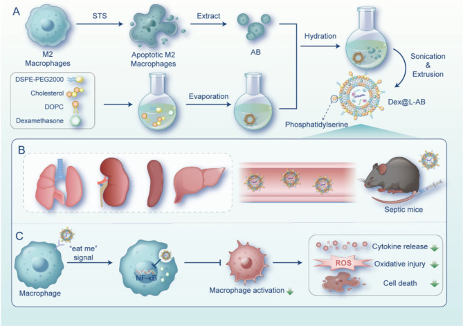
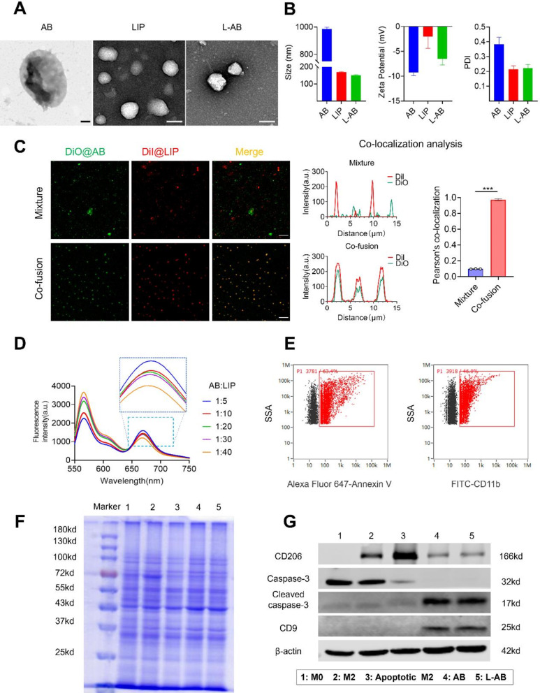
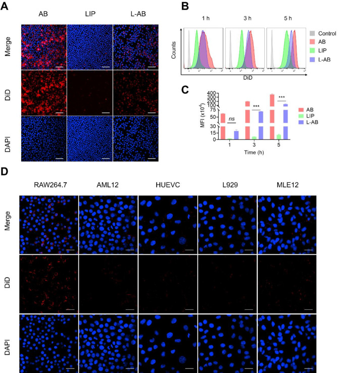
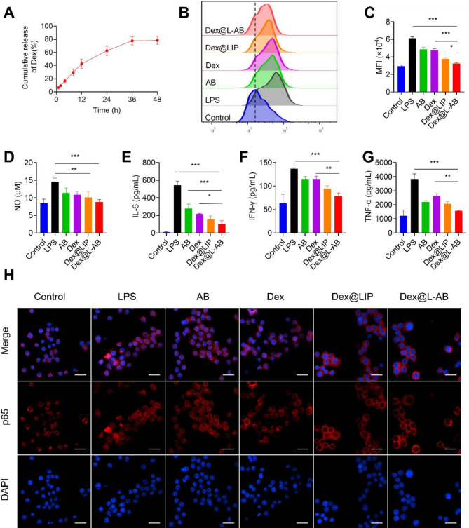
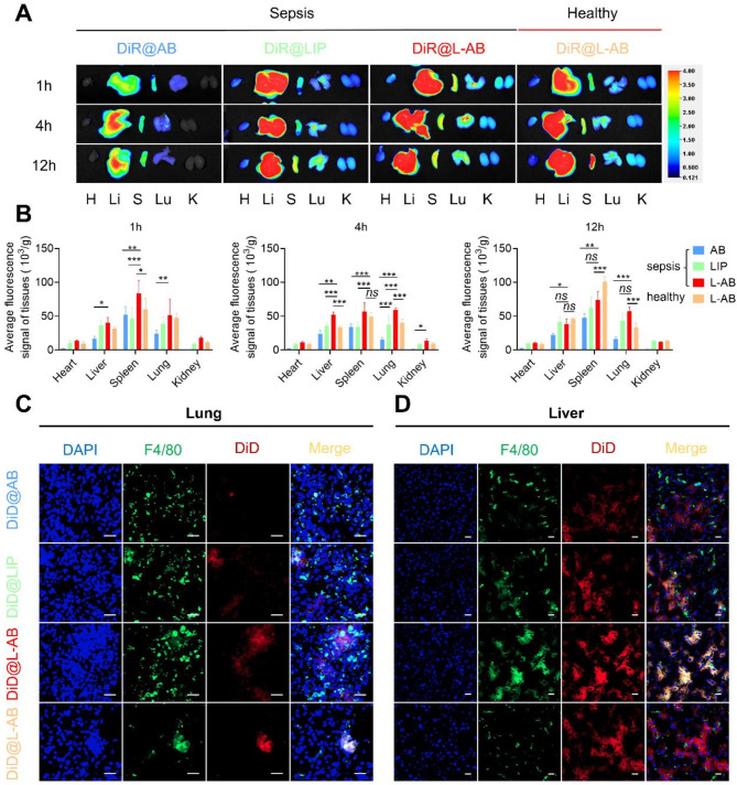
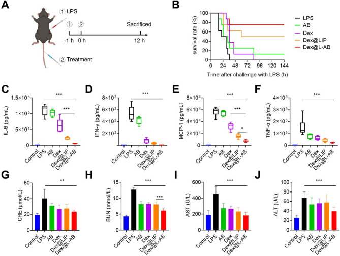
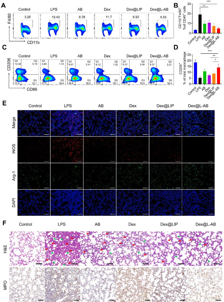
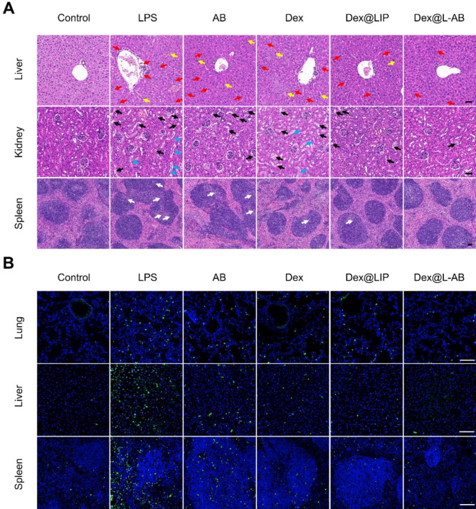
Sepsis is a severe immune response to pathogens that is associated with high mortality rate and a paucity of efficacious treatment options. It is characterized by the hyperactivation of macrophages and the occurrence of cytokine storms. Given the anti-inflammatory properties of M2 macrophages and their derived apoptotic bodies (AB), as well as the specific uptake of these by macrophages, a novel approach was employed to combine AB with artificial liposomes to create apoptotic body based biomimetic hybrid nanovesicles (L-AB). The L-AB effectively inherited "eat me" signaling molecules on the surface of the AB, thereby facilitating their targeted uptake by macrophages in both in vitro and in vivo settings. The administration of L-AB for the delivery of dexamethasone effectively augmented the therapeutic efficacy of the drug, mitigated macrophage hyperactivation and tissue damage in vivo, and consequently enhanced the survival rate of septic mice. Taken together, these findings suggest that the apoptotic body biomimetic nanovesicles may represent a potential drug delivery system capable of specifically targeting macrophages for the treatment of sepsis.

Keywords: Apoptotic body; Biomimetic carrier; Cytokine storm; Macrophages; Sepsis.


Conflict of interest statement

Declarations. Ethics approval and consent to participate: All the animal experiments were approved by the Experimental Animal Center of HUST. Animal experiments were carried out in compliance with the Guide for the Animals Care and Ethics Committee of HUST, with the assigned [2023] IACUC number (3678). Consent for publication: All authors consent for publication. Competing interests: The authors declare no competing interests.

Figures

Scheme 1
Scheme 1
Schematic illustration of apoptotic body based biomimetic hybrid nanovesicles for sepsis therapy. (A) Scheme of Dex@L-AB preparation process. (B) Dex@L-AB distributed to the major organs of septic mice through blood circulation after intravenous injection. (C) Upon recognition by macrophages through the “eat me” signal, Dex@L-AB is internalized and released into the cells to inhibit NF- κB pathway, thereby suppressing macrophage activation and reducing cytokine release, oxidative damage, and cell death. Painted by FigDraw
Fig. 1
Fig. 1
Preparation and characterization of L-AB. (A) TEM images of AB, LIP and L-AB. Scale bar = 200 nm. (B) Hydrodynamic size distribution and ζ potential of AB, LIP, and L-AB (n = 3). (C) CLSM images and co-localization analysis of a physical mixture (LIP with AB) (mixture) and L-AB (co-fusion) (red, LIP-DiI; green, AB-DiO; Scale bar = 10 μm). (D) FRET results of L-AB with different fusion ratios of LIP and AB. (E) The expression of PS and CD11b on the L-AB surface which was detected by nanoflow cytometry. (F) SDS-PAGE images of marker, 1: M0; 2: M2; 3: apoptotic M2; 4: AB; 5: L-AB.(G) Western blot of specific makers of the M0/M2/apoptotic M2 cell, AB and L-AB, 1: M0; 2: M2; 3: apoptotic M2; 4: AB; 5: L-AB
Fig. 2
Fig. 2
Cellular uptake of L-AB. (A) Fluorescence images of cellular uptake of AB, LIP, and L-AB on RAW264.7. Scale bar = 50 μm. (B, C) FCM quantification of the in vitro cellular uptake of AB, LIP, and L-AB by RAW264.7 (n = 3). (D) Cellular uptake of L-AB in different cells. Scale bar = 25 μm. Data are displayed as mean ± SD. ***P < 0.001, ns., not significant
Fig. 3
Fig. 3
In vitro anti-inflammatory and antioxidant activities of Dex@L-AB. (A) Release profiles of Dex@L-AB (n = 3). (B, C) Quantitative detection and analysis of intracellular ROS levels by FCM on RAW264.7 (n = 3). (D) NO from the supernatant of inflamed macrophages after incubation with various treatments (n = 3). (E-G) Cytokine levels of IL-6, IFN-γ, and TNF-α from the supernatant of inflamed macrophages after incubation with various treatments (n = 3). (H) Detection of p65 nuclear translocation via immune-fluorescence staining, scale bar = 25 μm. Data are displayed as mean ± SD. **P < 0.01, ***P < 0.001
Fig. 4
Fig. 4
Ex vivo biodistribution of L-AB. (A) Ex vivo biodistribution of DiR@AB, DiR@LIP, and DiR@L-AB in main organs at different time intervals via IVIS imaging. (B) Quantitative analysis of the DiR fluorescence signal in septic mice or healthy mice after intravenous injection of DiR@AB, DiR@LIP, and DiR@L-AB (n = 3). H, Li, S, Lu, and K represent heart, liver, spleen, lung, and kidney, respectively. (C, D) The localization of DiD@AB, DiD@LIP, and DiD@L-AB within the lung and liver, immunofluorescence analysis of lung and liver sections stained with F4/80. The sections were visualized using blue (DAPI), green (F4/80), and red (DiD) fluorescence, scale bar = 20 μm. Data are displayed as mean ± SD. *P < 0.05, **P < 0.01, ***P < 0.001
Fig. 5
Fig. 5
Therapeutic effects of Dex@L-AB in vivo. (A) Experimental procedures for the LPS-induced sepsis mouse model. (B) Survival rate of mice in different groups (n = 7). (C-F) The levels of proinflammatory cytokines IL-6, IFN-γ, MCP-1 and TNF-α in serum (n = 5). (G-J) Blood biochemistry analysis of CRE, BUN, ALT and AST (n = 5). Data are displayed as mean ± SD. *P < 0.05, **P < 0.01, ***P < 0.001
Fig. 6
Fig. 6
Therapeutic effect for experiencing sepsis-induced acute lung injury. (A) Representative plots of CD11b+ F4/80+ cells as a percentage of the total CD45+ cells population and (B) corresponding quantification results after various treatments (n = 5). (C) Representative plots of CD206+ cells as a percentage of the total macrophages population and (D) corresponding quantification results after various treatments (n = 5). (E) Representative fluorescence images of iNOS and Arg-1 in lung tissue after different treatments. (F) H&E staining and IHC staining of MPO of lung after different treatments (red arrows indicate inflammatory cells infiltration, green arrows indicate thickened alveolar walls). Scale bar = 100 μm. Data are displayed as mean ± SD. *P < 0.05, **P < 0.01, ***P < 0.001
Fig. 7
Fig. 7
Dex@L-AB relieved tissue injuries and cellular apoptosis of septic mice. (A) H&E staining of liver, kidney and spleen tissues. Red arrows indicate inflammatory cells infiltration; yellow arrows indicate liver necrosis cells; black arrows indicate vacuolated renal tubules; blue arrows indicate necrotic shedding of renal tubular epithelial cells to form casts and white arrows indicate disorganized germinal centers. (B) TUNEL staining of lung, liver and spleen tissues (green fluorescence: TUNEL positive cells; blue fluorescence: cell nucleus). Scale bar = 100 μm

References

    1. Cecconi M, Evans L, Levy M, Rhodes A. Sepsis and septic shock. Lancet. 2018;392(10141):75–87. - PubMed
    1. Singer M, Deutschman CS, Seymour CW, Shankar-Hari M, Annane D, Bauer M, Bellomo R, Bernard GR, Chiche J-D, Coopersmith CM, et al. The third international consensus definitions for sepsis and septic shock (Sepsis-3). JAMA. 2016;315(8):801–10. - PMC - PubMed
    1. Coperchini F, Chiovato L, Croce L, Magri F, Rotondi M. The cytokine storm in COVID-19. An overview of the involvement of the chemokine/chemokine-receptor system. Cytokine Growth Factor Rev. 2020;53:25–32. - PMC - PubMed
    1. Zheng X, Xing YJ, Sun K, Jin HZ, Zhao W, Yu F. Combination therapy with resveratrol and celastrol using folic acid-functionalized exosomes enhances the therapeutic efficacy of sepsis. Adv Healthc Mater. 2023;12(27):2301243. - PubMed
    1. Lee J, Son W, Hong J, Song Y, Yang CS, Kim YH. Down-regulation of TNF-α via macrophage-targeted RNAi system for the treatment of acute inflammatory sepsis. J Controlled Release. 2021;336:344–53. - PubMed

MeSH terms

LinkOut - more resources