. 2024 Dec 19;14(1):33.
doi: 10.1186/s13395-024-00366-y.

Regulation of injury-induced skeletal myofiber regeneration by glucose transporter 4 (GLUT4)

Affiliations

Regulation of injury-induced skeletal myofiber regeneration by glucose transporter 4 (GLUT4)

Tyler J Sermersheim et al. Skelet Muscle. .

Abstract

Background: Insulin resistance and type 2 diabetes impair cellular regeneration in multiple tissues including skeletal muscle. The molecular basis for this impairment is largely unknown. Glucose uptake via glucose transporter GLUT4 is impaired in insulin resistance. In healthy muscle, acute injury stimulates glucose uptake. Whether decreased glucose uptake via GLUT4 impairs muscle regeneration is presently unknown. The goal of this study was to determine whether GLUT4 regulates muscle glucose uptake and/or regeneration following acute injury.

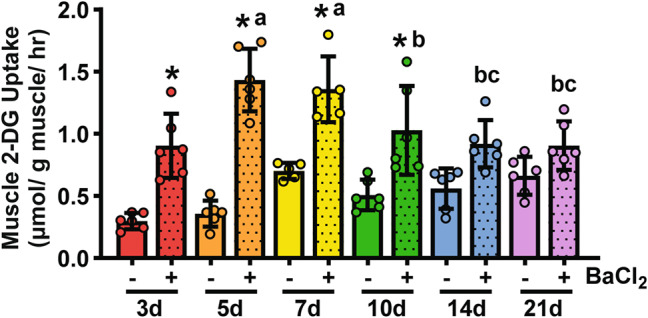
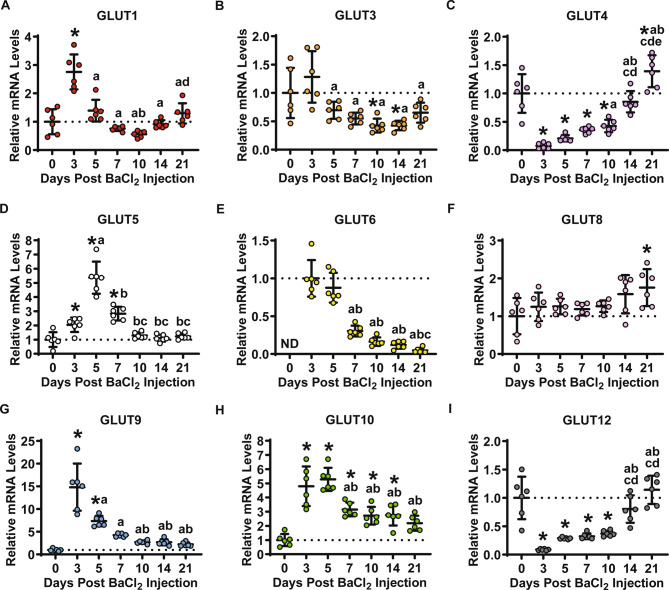
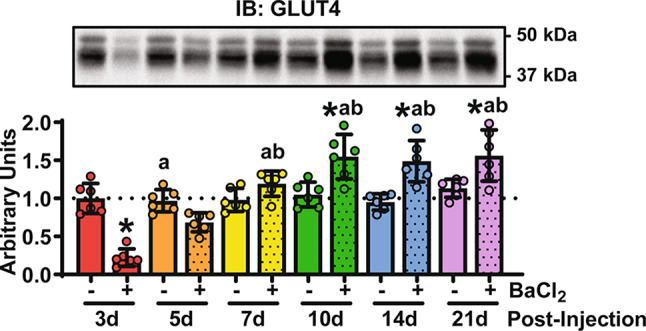
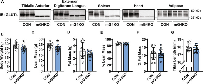
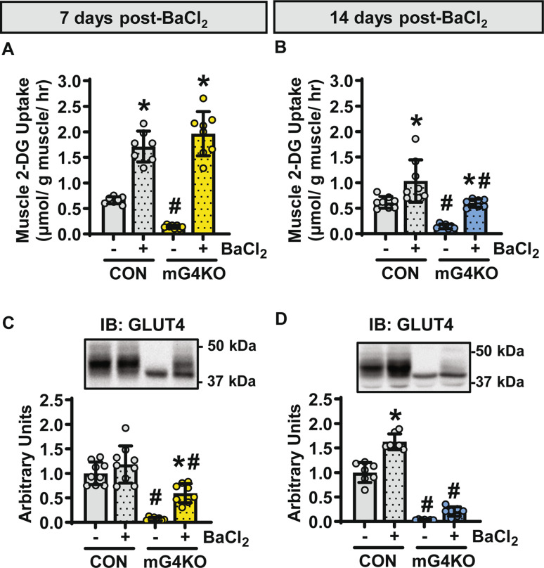
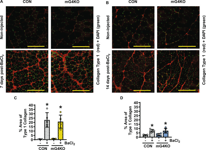
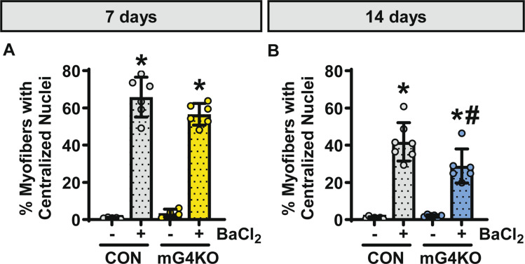
Methods: Tibialis anterior and extensor digitorum longus muscles from wild-type, control, or muscle-specific GLUT4 knockout (mG4KO) mice were injected with the myotoxin barium chloride to induce muscle injury. After 3, 5, 7, 10, 14, or 21 days (in wild-type mice), or after 7 or 14 days (in control & mG4KO) mice, muscles were isolated to examine [3H]-2-deoxyglucose uptake, GLUT4 levels, extracellular fluid space, fibrosis, myofiber cross-sectional area, and myofiber centralized nuclei.

Results: In wild-type mice, muscle glucose uptake was increased 3, 5, 7, and 10 days post-injury. There was a rapid decrease in GLUT4 protein levels that were restored to baseline at 5-7 days post-injury, followed by a super-compensation at 10-21 days. In mG4KO mice, there were no differences in muscle glucose uptake, extracellular fluid space, muscle fibrosis, myofiber cross-sectional areas, or percentage of centrally nucleated myofibers at 7 days post-injury. In contrast, at 14 days injured muscles from mG4KO mice exhibited decreased glucose uptake, muscle weight, myofiber cross sectional areas, and centrally nucleated myofibers, with no change in extracellular fluid space or fibrosis.

Conclusions: Collectively, these findings demonstrate that glucose uptake via GLUT4 regulates skeletal myofiber regeneration following acute injury.

Keywords: Barium chloride; Diabetes mellitus type 2; Extracellular fluid; Fibrosis; Glucose; Glucose transporter type 4; Insulin resistance; Knockout; Mice; Muscle; Regeneration.


Conflict of interest statement

Declarations. Ethical approval: Procedures were performed in accordance with the Indiana University School of Medicine Institutional Animal Care and Use Committee (IACUC, protocol# 21053 approved on 17 May 2021 and protocol# 21118 approved on 31 August 2021), and the National Institutes of Health Guidelines for the Care and Use of Laboratory Animals. Consent for publication: Not applicable. Competing interests: The authors declare no competing interests.

Figures

Fig. 1
Fig. 1
BaCl2-induced muscle injury stimulates a rapid and sustained increase in muscle glucose uptake. In male, wild-type C57BL/6J mice, 50 µl of a 1.2% BaCl2 solution was injected into the extensor digitorum longus (EDL) muscles of one leg. The contralateral leg was not injected. After 3, 5, 7, 10, 14 or 21 days, mice were fasted overnight, anesthetized with pentobarbital sodium, and euthanized. EDL muscles were incubated in [3H]-2-deoxyglucose to assess muscle glucose uptake. Data are presented as individual data points plus the mean ± standard deviation, with N = 5–6 muscles/group. Statistical significance was defined as P < 0.05, determined by Two-Way ANOVA with Tukey’s posthoc analysis, and denoted by ‘*’ vs. non-injected control muscle (-), ‘a’ vs. 3 days, ‘b’ vs. 5 days, and ‘c’ vs. 7 days
Fig. 2
Fig. 2
BaCl2-induced injury stimulates time-dependent alterations in the expression of multiple glucose transporters in skeletal muscle. In male, wild-type C57BL/6J mice, 50 µl of a 1.2% BaCl2 solution was injected into the tibialis anterior (TA) muscles of one leg. The contralateral leg was not injected (Day 0 muscles). After 3, 5, 7, 10, 14 or 21 days, TA muscles were collected and RNA isolated to assess glucose transporter (GLUT) levels by RT-qPCR. A: GLUT1; B: GLUT3; C: GLUT4; D: GLUT5; E: GLUT6; F: GLUT8; G: GLUT9; H: GLUT10; and I: GLUT12. Data are presented as individual data points plus the mean ± standard deviation, with N = 6 muscles/group. Statistical significance was defined as P < 0.05, determined by One-Way ANOVA with Tukey’s posthoc analysis, and denoted by ‘*’ vs. non-injected control muscle, ‘a’ vs. 3 days, ‘b’ vs. 5 days, ‘c’ vs. 7 days, ‘d’ vs. 10 days, and ‘e’ vs. 14 days
Fig. 3
Fig. 3
BaCl2-induced muscle injury stimulates a time-dependent alteration in muscle GLUT4 protein levels. In male, wild-type C57BL/6J mice, 50 µl of a 1.2% BaCl2 solution was injected into the tibialis anterior (TA) muscles of one leg. The contralateral leg was not injected. After 3, 5, 7, 10, 14 or 21 days, TA muscles were collected and protein extracted to assess total muscle GLUT4 protein levels by immunoblot (IB) analyses. Data are presented as individual data points plus the mean ± standard deviation, with N = 6 muscles/group. Statistical significance was defined as P < 0.05, determined by Two-Way ANOVA with Tukey’s posthoc analysis, and denoted by ‘*’ vs. non-injected control muscle (-), ‘a’ vs. 3 days, and ‘b’ vs. 5 days
Fig. 4
Fig. 4
Body weight and body composition are impaired in muscle-specific GLUT4 knockout mice. All measures were made in 11–12 week old, male control (CON) and muscle-specific GLUT4 knockout (mG4KO) mice. (A) Tissues were collected to assess GLUT4 protein levels by immunoblot (IB) analysis. Images from N = 3 mice/group. (B) Body weight. (Panels C-F) Body composition was assessed by EchoMRI™. (C) Lean mass. (D) Fat mass. (E) Percent lean mass relative to body weight. (F) Percent fat mass relative to body weight. (G) Tibias were isolated and tibia length measured with a micrometer. Data are presented as individual data points plus the mean ± standard deviation, with N = 10–13 mice/group. Statistical significance was defined as P < 0.05, determined by t-tests, and denoted by ‘*’ vs. CON mice
Fig. 5
Fig. 5
Muscle-specific deletion of GLUT4 impairs muscle glucose uptake at 14 days post BaCl2-induced injury. In 13–14 week old, male control (CON) and muscle-specific GLUT4 knockout (mG4KO) mice, BaCl2 solution was injected into the muscles of one leg. The contralateral leg was not injected. Muscles were examined at 7 days or 14 days post-injury. (Panels A + B) Extensor digitorum longus muscles were incubated in [3H]-2-deoxyglucose to assess glucose uptake. (Panels C + D) Tibialis anterior muscles were frozen to assess GLUT4 protein levels by immunoblot (IB) analyses. Representative images and quantification of band intensity. Data are presented as individual data points plus the mean ± standard deviation, with N = 7–9 muscles/group. Statistical significance was defined as P < 0.05, determined by Two-Way ANOVA with Tukey’s posthoc analysis, and denoted by ‘*’ vs. non-injected control muscle (-), and ‘#’ vs. CON mice
Fig. 6
Fig. 6
Muscle-specific deletion of GLUT4 reduces muscle weight but not extracellular fluid space post BaCl2-induced injury. In 13–14 week old, male control (CON) and muscle-specific GLUT4 knockout (mG4KO) mice, BaCl2 solution was injected into the muscles of one leg. The contralateral leg was not injected. Extensor digitorum longus (EDL) muscles were examined at 7 days (panels A + C) or 14 days (panels B + D) post-injury. (A + B) EDL muscles were excised and immediately weighed (N = 11–14 muscles/group). (C + D) EDL muscles were incubated in [14C]-mannitol to assess extracellular fluid space (N = 7–8 muscles/group). Data are presented as individual data points plus the mean ± standard deviation. Statistical significance was defined as P < 0.05, determined by Two-Way ANOVA with Tukey’s posthoc analysis, and denoted by ‘*’ vs. non-injected control muscle (-), and ‘#’ vs. CON mice
Fig. 7
Fig. 7
Muscle-specific deletion of GLUT4 does not alter muscle fibrosis post BaCl2-induced injury. In 13–14 week old, male control (CON) and muscle-specific GLUT4 knockout (mG4KO) mice, BaCl2 solution was injected into the muscles of one leg. The contralateral leg was not injected. (Panels A + B) Representative images of tibialis anterior muscle cross-sections immunolabeled for type 1 collagen (red) and stained with DAPI to label nuclei (green) for the (A) 7 day post-BaCl2 injured muscles and for the (B) 14 day post-BaCl2 injured muscles. Scale bar = 200 μm. (Panels C + D) Quantification of the percent area of type 1 collagen in the muscles from the (C) 7 day post-BaCl2 injected mice (N = 4–5 muscles/group), and (D) 14 day post-BaCl2 injected mice (N = 4–6 muscles/group). Data are presented as individual data points plus the mean ± standard deviation. Statistical significance was defined as P < 0.05, determined by Two-Way ANOVA with Tukey’s posthoc analysis, and denoted by ‘*’ vs. non-injected control muscle (-)
Fig. 8
Fig. 8
Muscle-specific deletion of GLUT4 impairs myofiber size at 14 days post BaCl2-induced injury. In 13–14 week old, male control (CON) and muscle-specific GLUT4 knockout (mG4KO) mice, BaCl2 solution was injected into the muscles of one leg. The contralateral leg was not injected. (A + B) Representative images of muscle cross-sections with the myofiber cell perimeter immunolabeled for laminin (violet) and the nuclei stained with DAPI (green) for the (A) 7 day post-BaCl2 injured muscles, and for the (B) 14 day post-BaCl2 injured muscles. Scale bar = 200 μm. (Panels C-J) Quantification of the average myofiber cross-sectional areas (CSA) and percent frequency of myofibers with CSAs divided into groups of 450–500 um2. (C + D & G + H) Data from mice euthanized 7 days after BaCl2 injections (N = 4–6 muscles/group). (E + F & I + J) Data from mice euthanized 14 days after BaCl2 injections (N = 6–7 muscles/group). Data are presented as individual data points plus the mean ± standard deviation, or as mean ± standard deviation. Statistical significance was defined as P < 0.05, determined by t-tests, and denoted by ‘*’ vs. non-injected control muscle (-)
Fig. 9
Fig. 9
Muscle-specific deletion of GLUT4 reduces the number of centrally nucleated myofibers post BaCl2-induced injury. In 13–14 week old, male control (CON) and muscle-specific GLUT4 knockout (mG4KO) mice, BaCl2 solution was injected into the muscles of one leg. The contralateral leg was not injected. Quantification of the percentage of myofibers with centralized nuclei in muscles from mice euthanized at (A) 7 days (N = 4–6 muscles/group), and (B) 14 days post-injection (N = 6–7 muscles/group). Data are presented as individual data points plus the mean ± standard deviation. Statistical significance was defined as P < 0.05, determined by Two-Way ANOVA with Tukey’s posthoc analysis, and denoted by ‘*’ vs. non-injected control muscle (-), and ‘#’ vs. CON mice

References

    1. Nguyen MH, Cheng M, Koh TJ. Impaired muscle regeneration in ob/ob and db/db mice. ScientificWorldJournal. 2011;11:1525–35. - PMC - PubMed
    1. D’Souza DM, Trajcevski KE, Al-Sajee D, Wang DC, Thomas M, Anderson JE et al. Diet-induced obesity impairs muscle satellite cell activation and muscle repair through alterations in hepatocyte growth factor signaling. Physiol Rep. 2015;3. - PMC - PubMed
    1. Hu Z, Wang H, Lee IH, Modi S, Wang X, Du J, et al. PTEN inhibition improves muscle regeneration in mice fed a high-fat diet. Diabetes. 2010;59:1312–20. - PMC - PubMed
    1. Baron AD, Kolterman OG, Bell J, Mandarino LJ, Olefsky JM. Rates of noninsulin-mediated glucose uptake are elevated in type II diabetic subjects. J Clin Investig. 1985;76:1782–8. - PMC - PubMed
    1. Baron AD, Brechtel G, Wallace P, Edelman SV. Rates and tissue sites of non-insulin- and insulin-mediated glucose uptake in humans. Am J Physiol. 1988;255:E769–74. - PubMed

LinkOut - more resources