. 2024 Dec 18;10(1):96.
doi: 10.1186/s40942-024-00612-x.

Clinical characteristics and risk factors of bacillary layer detachment in central serous chorioretinopathy: a comparative multicenter study

Affiliations

Clinical characteristics and risk factors of bacillary layer detachment in central serous chorioretinopathy: a comparative multicenter study

Antonio M Casella et al. Int J Retina Vitreous. .

Abstract

Background: Central serous chorioretinopathy (CSC) is marked by serous retinal detachments caused by fluid leakage from the retinal pigment epithelium, often associated with stress, psychiatric disorders and the use of corticosteroids. This study aims to investigate the clinical and systemic characteristics associated with BALAD in patients with CSC, comparing those with and without BALAD to clarify its function as a biomarker of CSC severity and improve diagnostic and treatment approaches.

Purpose: Compare the clinical characteristics, risk factors, and optical coherence tomography (OCT) findings in patients with Central Serous Chorioretinopathy (CSC) with and without Bacillary Layer Detachment (BALAD), and to identify the distinguishing features and associated conditions of CSC with BALAD.

Methods: This observational, retrospective, multicenter case-control study collected data from 12 retina centers worldwide on patients with central serous chorioretinopathy (CSC) from December 1, 2022, to April 1, 2023. CSC was defined by serous retinal detachment and fluid leakage through the retinal pigment epithelium. Patients underwent detailed evaluations, including OCT, and were classified as having acute or chronic CSC. Inclusion criteria included a CSC diagnosis with RPE leakage, BALAD confirmed by three authors, age over 18, and a detailed medical history from the 30 days before symptom onset. The study assessed visual acuity, choroidal thickness, psychiatric disorders, corticosteroid use, prior CSC treatments, and hyperreflective material on OCT.

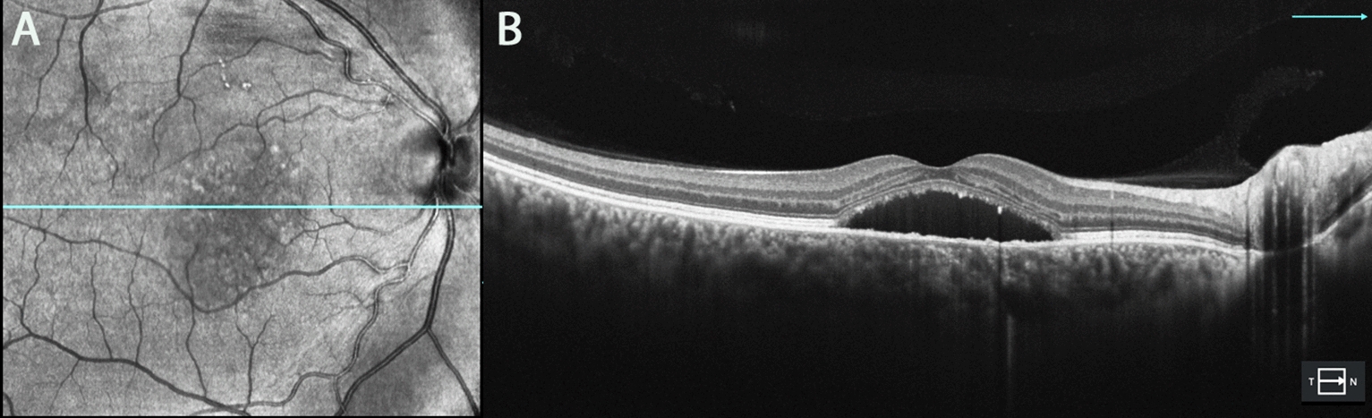
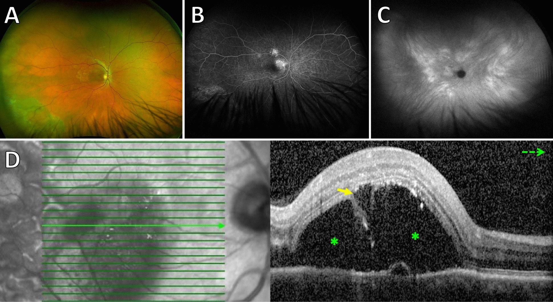
Results: Thirty-seven patients (40 eyes; mean age, 48.0 ± 11.9 years) had CSC and BALAD and were followed for a mean of 4.92 ± 6.65 months. The control group was comprised of 40 patients with CSC without BALAD (40 eyes; mean age, 48.2 ± 11.9 years). On clinical examination, BALAD was as a circular, yellowish macular lesion. On OCT, BALAD was a detachment of the ellipsoid zone with splitting of the photoreceptor inner segment. BALAD was associated with psychiatric disorders (p = 0.014), use of corticosteroids (p = 0.004), previous treatment for CSC (p = 0.041) and thickened choroid (p = 0.036).

Conclusions: BALAD in CSC differs from a typical CSC due to the presence of a circular, yellowish macular lesion, detachment of the ellipsoid zone, segmentation of the inner segment of the photoreceptor, a thicker choroid, the use of corticosteroids, and generally more aggressive previous treatments. These results suggest that BALAD may serve as a valuable biomarker for the severity of CSC and highlight the influence of inflammation and previous treatments.

Keywords: Bacillary layer detachment; Central serous chorioretinopathy; Inflammation; Photodynamic therapy.


Conflict of interest statement

Declarations. Ethics approval and consent to participate: This study was performed in line with the principles of the Declaration of Helsinki. Approval was granted by all local ethics committee at each participating center. Consent for publication: Written informed consent was obtained from all patients in the study. Competing interests: AMC is editor-in-chief for IJRV, but he is not partipating of the peer review process.

Figures

Fig. 1
Fig. 1
Optical coherence tomography images (A, B) of a 54-year-old patient in the control group with chronic CSC
Fig. 2
Fig. 2
Optical coherence tomography (A, B) of a patient with chronic CSC and BALAD. Patient had low vision shortly after receiving treatment with photodynamic therapy. The yellow arrow represents BALAD, and the green asterisk indicates SRF. BALAD bacillary layer detachments, SRF subretinal fluid
Fig. 3
Fig. 3
Fundus photograph (A), fluorescein angiography (B), indocyanine green angiography (C) and optical coherence tomography (D) of a 58-year-old male patient who developed BALAD after treatment of chronic CSC by photodynamic therapy. Optical coherence tomography angiography (OCTA) excluded choroidal neovascularization. The yellow arrow represents BALAD, and the green asterisk indicates SRF. BALAD bacillary layer detachments, SRF subretinal fluid
Fig. 4
Fig. 4
Fundus photograph (A), Autofluorescence (B), and optical coherence tomography images (C and D) of the left eye of a 26-year-old man with acute bilateral central serous chorioretinopathy with BALAD. Images show the presence of hyper-reflectivity within the BALAD cavity (red arrow). The yellow arrow represents BALAD, and the green asterisk indicates SRF. The red arrow represents materials with hyperreflectivity. BALAD bacillary layer detachments, SRF subretinal fluid
Fig. 5
Fig. 5
Sequence of OCT images of a 43-year-old woman with acute CSC and BALAD in the right eye. A 20 days after COVID-19 diagnosis and corticosteroid use. B 30-day follow-up after stopping corticosteroids. C 60-day follow-up showing resolution. The yellow arrow indicates BALAD, and the green asterisk represents subretinal fluid (SRF). The yellow arrow represents BALAD, and the green asterisk indicates SRF. BALAD bacillary layer detachments, SRF Subretinal fluid
Fig. 6
Fig. 6
Fundus photographs (A, C) and optical coherence tomography (OCT) images (B, D) of a 22-year-old male who presented with low vision 28 days after developing acute central serous chorioretinopathy (CSC), following treatment with high doses of oral corticosteroids for systemic lupus erythematosus. It is likely that corticosteroid use exacerbated the serous retinal detachments, leading to vascular compromise that affects the integrity of the retinal pigment epithelium (RPE) and neurosensory retina, promoting the onset of CSC. Images (C) and (D) show the patient at a 30-day follow-up. The yellow arrow indicates Bacillary Layer Detachment (BALAD), and the green asterisk marks subretinal fluid (SRF). BALAD Bacillary Layer Detachments, SRF Subretinal Fluid

References

    1. Yannuzzi LA. Central serous chorioretinopathy: a personal perspective. Am J Ophthalmol. 2010;149(3):361–3. 10.1016/j.ajo.2009.11.017. - PubMed
    1. Spaide RF, Campeas L, Haas A, et al. Central serous chorioretinopathy in younger and older adults. Ophthalmology. 1996;103:2070–9. 10.1016/s0161-6420(96)30386-2. - PubMed
    1. Ramtohul P, Engelbert M, Malclès A, et al. Bacillary layer detachment: multimodal imaging and histologic evidence of a novel optical coherence tomography terminology: literature review and proposed theory. Retina. 2021;41:2193–207. 10.1097/IAE.0000000000003217. - PubMed
    1. Liu XY, Peng XY, Wang S. Features of optical coherence tomography for the diagnosis of Vogt-Koyanagi-Harada disease. Retina. 2016;36:2116–23. 10.1097/IAE.0000000000001076. - PubMed
    1. Rajesh B, Kaur A, Giridhar A, Gopalakrishnan M. “Vacuole” sign adjacent to retinal pigment epithelial defects on spectral domain optical coherence tomography in central serous chorioretinopathy associated with subretinal fibrin. Retina. 2017;37:316–24. 10.1097/IAE.0000000000001192. - PubMed

LinkOut - more resources