Review
. 2024 Dec 18;29(1):155.
doi: 10.1186/s11658-024-00671-w.

Decoding the enigmatic estrogen paradox in pulmonary hypertension: delving into estrogen metabolites and metabolic enzymes

Affiliations
Review

Decoding the enigmatic estrogen paradox in pulmonary hypertension: delving into estrogen metabolites and metabolic enzymes

Qiang You et al. Cell Mol Biol Lett. .

Abstract

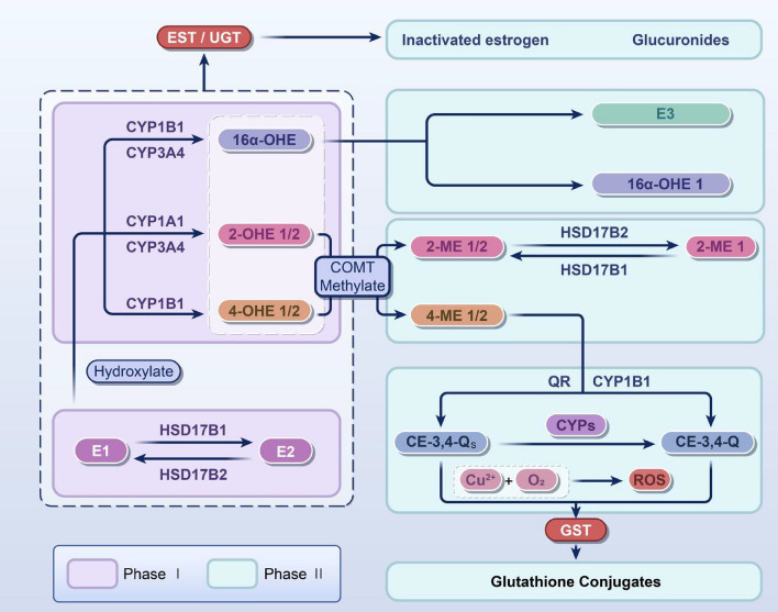
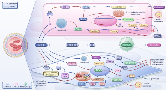
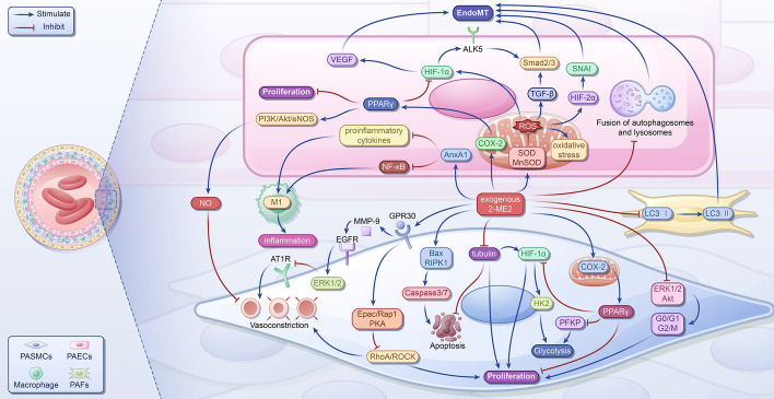
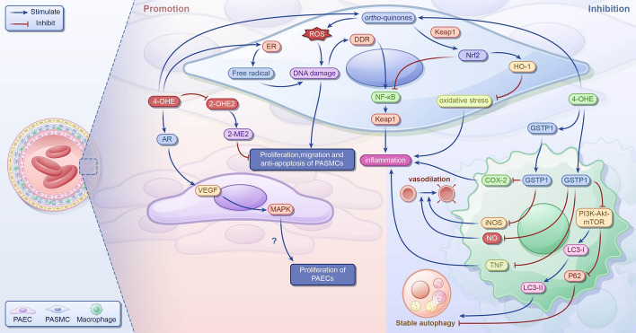
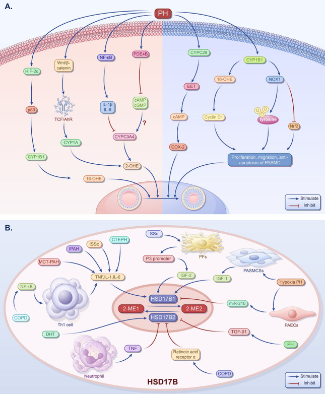
Pulmonary hypertension (PH) presents a puzzling sex bias, being more prevalent in women yet often less severe than in men, and the underlying reasons remain unclear. Studies using animal models, and limited clinical data have revealed a protective influence of exogenous estrogens, known as the estrogen paradox. Research suggests that beyond its receptor-mediated effects, estrogen acts through metabolites such as 2-ME2, 4-OHE2, and 16-OHE2, which are capable of exhibiting protective or detrimental effects in PH, prompting the need to explore their roles in PH to untangle sex differences and the estrogen paradox. Hypoxia disrupts the balance of estrogen metabolites by affecting the enzymes responsible for estrogen metabolism. Delving into the role of these metabolic enzymes not only illuminates the sex difference in PH but also provides a potential rationale for the estrogen paradox. This review delves into the intricate interplay between estrogen metabolites, metabolic enzymes, and PH, offering a deeper understanding of sex-specific differences and the perplexing estrogen paradox in the context of this condition.

Keywords: CYPs; Estrogen; Estrogen metabolites; HSD17B; Hypoxia; Pulmonary hypertension.


Conflict of interest statement

Declarations. Ethics approval and consent to participate.: Not applicable. Consent for publication: Not applicable. Competing interests: The authors declare that there is no conflict of interest.

Figures

Fig. 1
Fig. 1
Main metabolic processes of estrogen. The main metabolic process of estrogen includes two stages. The first stage mainly forms metabolites dominated by 2-OHE, 4-OHE, and 16aα-OHE through CYPs. The second phase involves further metabolic processes of hydroxylated metabolites, including three major pathways: (1) methylation: COMT can convert 2-OHE and 4-OHE into 2-ME1/2 and 4-ME1/2; (2) glucuronidation: UGT/EST can convert estrogen or its hydroxylated metabolites into glucuronate; and (3) sulfonation: GST can convert these two quinone metabolites into less toxic small molecules, such as glutathione conjugates. E1, estrone; E2, estradiol; E3, estriol; EST, estrogen sulfotransferase; UGT, UDP-glucuronosyltransferase; 16α-OHE 1, 16α-hydroxyestrone; 16α-OHE, 16α-hydroxyestradiol; 2-OHE 1/2, 2-hydroxyestrone/2-hydroxyestradiol; 4-OHE 1/2, 4-hydroxyestrone/4-hydroxyestradiol; 2-ME2 1/2, 2-methoxyestrone/2-methoxyestradiol; 4-ME 1/2, 4-methoxyestrone/4-methoxyestradiol; QR, quinone reductase; CYPs, cytochrome p450s; CE-3:4-QS, catechol estrogen-3:4-semiquinone; CE-3:4-Q, catechol estrogen-3:4-benzoquinone; GST, glutathione S-transferase; COMT, catechol O-methyltransferase; HSD17B, 17β-hydroxysteroid dehydrogenase; ROS, reactive oxygen species. The arrows in the figure indicate transformations
Fig. 2
Fig. 2
The mechanism of 16α-OHE promoting pulmonary hypertension. In PASMCs, 16α-OHE1 binds with ERα to inhibit Nrf2, resulting in upregulation of Nox1 and Nox4, decreased antioxidants increased ROS production, and irreversible PTP oxidation. Activation of the p38MARK pathway leads to increased phosphorylation of CRE region and ATF-2, upregulation of cyclinD1, and promotion of cell oxidative damage and proliferation. Upregulation of miRNA-29 inhibits expression of PPARγ, further reducing CD36 and Glut4 and upregulating PFKP, resulting in insulin resistance and increased aerobic glycolysis of 16α-OHE1, which may promote cell, migration, and antiapoptosis by reducing BMPR2 levels and inhibiting the BMPR2-Smad1/5/8-ID1, BMPR2-AkT-Wnt/β-Catenin, and BMPR2-PPARγ-apoe signaling pathways through lysosomal activation. In PAECs, 16α-OHE1 binds with Erα to inhibit X17, upregulate HIF-2α, and increases cyclin D2 and E2F1, promoting cell proliferation. Additionally, 16α-OHE1 inhibits PPARγ and attenuates mitochondrial bioenergy and insulin resistance, leading to metabolic abnormalities. Inhibition of PPARγ and BMPR2 promotes EndoMT through inhibition of p-Smad1/5/8-Smad4 signaling and enhancement of TGF-β-Smad2/3-Smad4 signaling. E3 stimulates the expression of TNF and IL-6, potentially exerting a proinflammatory effect in PH. E3, estriol; 16α-OHE1/2, 16α-hydroxyestrone/16α-hydroxyestradiol; TNF, tumor necrosis factor; IL-6, interleukin-6; Nrf2, nuclear factor E2-related factor 2; SOD1, superoxide dismutase 1; ROS, reactive oxygen species; Nox1/4, nicotinamide adenine dinucleotide phosphate oxidase1/4; PTP, protein tyrosine phosphatases; p38MARK, p38 mitogen-activated protein kinase; CRE, cAMP response element; ATF-2, activating transcription factor 2; ERs, estrogen receptors; SOX17, SRY-related HMG-box 17; HIF-2α, hypoxia-inducible factor 2α; E2F1, E2F transcription factor 1; PPARγ, peroxisome proliferator-activated receptor γ; CD36, cluster of differentiation 36; Glut4, glucose transporters type 4; PFKP, platelet-type phosphofructokinase; BMPR2, bone morphogenetic protein receptor type II; Id1, DNA binding 1; Akt, protein kinase B; apoE, apolipoprotein E; TGF-β, transforming growth factor-β; EndoMT, endothelial-to-mesenchymal transition; PASMC, pulmonary artery smooth muscle cell; PAEC, pulmonary arterial endothelial cell
Fig. 3
Fig. 3
The mechanism of 2-ME2 suppressing pulmonary hypertension. In PASMCs, 2-ME2 indirectly activates PPARγ by promoting COX-2 expression, inhibiting proliferation. It also binds GPR30, activating Epac/Rap1 and PKA, inhibiting RhoA/ROCK expression, and, ultimately, inhibiting proliferation and vasoconstriction. In addition, 2-ME2 inhibits the activation of ERK1/2/Akt, blocking the G0/G1 and G2/M phase of the cell cycle and inhibiting proliferation. It disrupts tubulin, promotes apoptosis, and activates PPARγ, inhibiting PFKP and HIF-1α/HK2 to inhibit glycolysis. Furthermore, 2-ME2 binds GPR30, transactivating EGFR by releasing MMP-9, to activate ERK1/2, inhibit AT1R expression, and inhibit vasoconstriction. In pulmonary artery endothelial cells (PAECs), 2-ME2 activates PPARγ, which may inhibit proliferation and activate PI3K/Akt/eNOS, increase NO release, and inhibit vasoconstriction. It also inhibits HIF-1α, VEGF, and TGF-β/Smad2/3 to inhibit EndoMT; 2-ME2 inhibits SOD and MnSOD to inhibit ROS and oxidative stress, inhibiting HIF1α, TGF-β, and HIF-2α/SNAI, ultimately inhibiting EndoMT. It also inhibits the fusion of autophagosomes and lysosomes to inhibit EndoMT. Additionally, 2-ME2 promotes AnxA1 expression, inhibiting proinflammatory cytokines and NF-κB and inhibiting inflammation; 2-ME2 inhibits macrophage activation, inflammation, and early LC3 transformation in PAFs to inhibit EndoMT. PPARγ, peroxisome proliferator-activated receptor γ; 2-ME2, 2-methoxyestradiol; Bax, Bcl-2-associated X protein; COX-2, cyclooxygenase-2; PFKP, platelet-type phosphofructokinase; HIF-1α, hypoxia-inducible factor-1α; HIF-2α, hypoxia-inducible factor-2α; HK2, hexokinase 2; RhoA, Ras homolog gene family member A; ROCK, Rho-kinase; ERK1/2, extracellular signal-regulated kinases1/2; GPR30, G protein-coupled receptor 30; Epac, exchange protein activated by cAMP; Rap1, Ras-associated protein 1; PKA, protein kinase A; Akt, protein kinase B; MMP-9, matrix metalloproteinase-9; EGFR, epidermal growth factor receptor; AT1R, angiotensin II type I receptor; PI3K, phosphatidylinositol 3-kinase; eNOS, endothelial nitric oxide synthase; NO, nitric oxide; VEGF, vascular endothelial-derived growth factor; ALK5, activin receptor‑like kinase-5; RIPK1, receptor-interacting protein kinase 1; SOD, superoxide dismutase; MnSOD, manganese superoxide dismutase; ROS, reactive oxygen species; TGF-β, transforming growth factor-β; SNAI, zinc finger protein SNAI family; AnxA1, annexin A1; NF-κB, nuclear factor-kappa B; LC3, light chain 3; EndoMT, endothelial-to-mesenchymal transition; PASMC, pulmonary artery smooth muscle cells; PAEC, pulmonary arterial endothelial cell
Fig. 4
Fig. 4
The role of 4-OHE in pulmonary hypertension; 4-OHEs competitively decrease the activity of 2-OHE2, inhibiting the formation of 2-ME2. They activate ERs, leading to the production of free radicals and generation of ortho-quinones, resulting in ROS production and DNA damage, which can cause dysfunction of PAECs and promote the proliferation and migration of PASMCs. By binding to ARs, 4-OHEs increase VEGF expression and activate ERK1/2, JNK, and p38MAPK pathways and promote the proliferation of AECs; 4-OHEs induce DDR and upregulate the levels of proinflammatory cytokines IL-1β, IL-6, and TNF, potentially aggravating the deterioration of PH. However, 4-OHEs also increase HO-1 levels through the Nrf2–Keap1–ARE pathway, inhibiting oxidative stress and PASMCs proliferation and promoting apoptosis. In lung tissue, they enhance Nrf2 expression, reducing the levels of proinflammatory cytokines IL-8 and TNF. In LPS-stimulated macrophages, 4-OHEs upregulate GSTP1, decreasing the levels proinflammatory cytokines TNF, NO, iNOS, and COX-2, mitigating inflammation, and potentially ameliorating PH deterioration. Furthermore, they inhibit the PI3K–Akt–mTOR pathway and LC3-II levels, decrease p62 levels, and enhance autophagy stability. 4-OHEs, 4-hydroxyestrogens; 2-OHE2, 2-hydroxyestradiol; 2-ME2, 2-methoxyestradiol; ERs, estrogen receptors; ARs, adrenergic receptors; VEGF, vascular endothelial-derived growth factor; ERK1/2, extracellular signal-regulated kinases1/2; JNK, c-Jun N-terminal kinase; p38MAPK, p38 mitogen-activated protein kinase; AECs, arterial endothelial cells; DDR, DNA damage response; IL, interleukin; TNF, tumor necrosis factor; HO-1, heme oxygenase 1; Nrf2, nuclear factor E2-related factor 2; Keap1, Kelch-like ECH-associated protein 1; ARE, antioxidant response element; LPS, lipopolysaccharide; COX-2, cyclooxygenase-2; GSTP1, glutathione S-transferase P1; NO, nitric oxide; iNOS, inducible nitric oxide synthase; PI3K, phosphatidylinositol 3-kinase; Akt, protein kinase B; mTOR, mechanistic target of rapamycin; LC3-II, light chain 3-II
Fig. 5
Fig. 5
The influence factors of CYPs and HSD17B in PH. A The influence factors of CYPs in pulmonary hypertension. In patients with PH or animal models, the expression of HIF-2α is increased, and the upregulation of p53 may promote the expression of CYP1B1 and increase the level of 16-OHE. The increased levels of IL-1β and IL-6 may inhibit the activity of CYP3A4 and decrease the level of 2-OHE. The activation of Wnt/β-catenin signaling pathway may promote the expression of CYP1A and increase the level of 2-OHE. The expression of PDE4B is increased leads to decreased levels of cAMP and cGMP, but the content of CYP3A is not clear. The increased expression of CYP2C29 and EET increases the expression of COX-2 through a cAMP-dependent pathway, thereby promoting endothelial cell proliferation and angiogenesis. The increased expression of CYP1B1 may promote the proliferation of PASMCs by increasing the levels of 16α-OHE and cyclin D1, promoting the expression of Nox1, stimulating the irreversible oxidation of protein tyrosine phosphatase, and reducing the activity of nuclear factor Nrf2 and the expression of its antioxidant genes. B The influence factors of HSD17B in PH. Under PH, OSoxidative stress activates the NF-κB channel and promotes of TNF, IL-6, and IL-1 by helper T cell type 1 45, which will upregulate HSD17B1. In addition, in PH, the upregulation of HSD17B1 and the increase of HSD17B1 will increase the production of E2 and decrease the production of E1. Due to TNF, ATRA, and DHT, HSD17B2 increases, resulting in increased E1 production and decreased E2 production. HIF-2α, hypoxia-inducible factor-2alpha; CYP, cytochrome P450; 2-OHE, 2-hydroxyestradiol; PH, pulmonary artery hypertension; OS, oxidative stress; IL, interleukin; PDE4B, phosphodiesterase 4B; cAMP, cyclic adenosine monophosphate; cGMP, cyclic guanosine 3′:5′-monophosphate; EET, endoscopic eradication therapy; MAP, mitogen-activated protein; COX-2, cyclooxygenase-2; Nox1, nicotinamide adenine dinucleotide phosphate oxidase1; Nrf2, nuclear factor E2-related factor 2; NF-κB, nuclear factor-kappa B; HSD17B, 17β-hydroxysteroid dehydrogenase; TNF, tumor necrosis factor; IGF, insulin-like growth factor

Similar articles

Cited by

References

    1. Mocumbi A, Humbert M, Saxena A, Jing ZC, Sliwa K, Thienemann F, Archer SL, Stewart S. Pulmonary hypertension. Nat Rev Dis Prim. 2024;10(1):1. 10.1038/s41572-023-00486-7. - PubMed
    1. Hansmann G. Pulmonary hypertension in infants, children, and young adults. J Am Coll Cardiol. 2017;69(20):2551–69. 10.1016/j.jacc.2017.03.575. - PubMed
    1. Dodson MW, Brown LM, Elliott CG. Pulmonary arterial hypertension. Heart Fail Clin. 2018;14(3):255–69. 10.1016/j.hfc.2018.02.003. - PubMed
    1. Rosenkranz S, Gibbs JS, Wachter R, De Marco T, Vonk-Noordegraaf A, Vachiery JL. Left ventricular heart failure and pulmonary hypertension. Eur Heart J. 2016;37(12):942–54. 10.1093/eurheartj/ehv512. - PMC - PubMed
    1. Brusca SB, Zou Y, Elinoff JM. How low should we go? Potential benefits and ramifications of the pulmonary hypertension hemodynamic definitions proposed by the 6th World Symposium. Curr Opin Pulm Med. 2020;26(5):384–90. 10.1097/MCP.0000000000000699. - PMC - PubMed