

Association between choroid plexus volume and cognitive function in community-dwelling older adults without dementia: a population-based cross-sectional analysis
- PMID: 39696504
- PMCID: PMC11654186
- DOI: 10.1186/s12987-024-00601-0
Association between choroid plexus volume and cognitive function in community-dwelling older adults without dementia: a population-based cross-sectional analysis
Abstract
Background: An increase in choroid plexus (CP) volume may be associated with cognitive decline in older individuals without dementia. In this study, we aimed to clarify whether CP volume can serve as an imaging marker of cognitive decline, determine how strongly CP volume is associated with cognitive decline, and explore factors associated with CP volume in older adults.
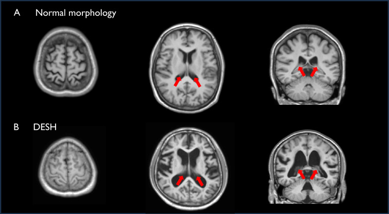
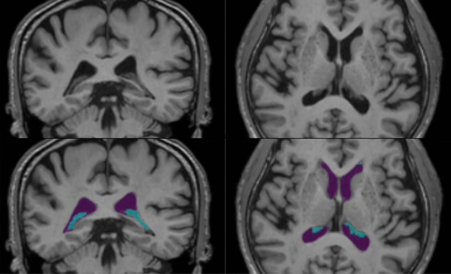
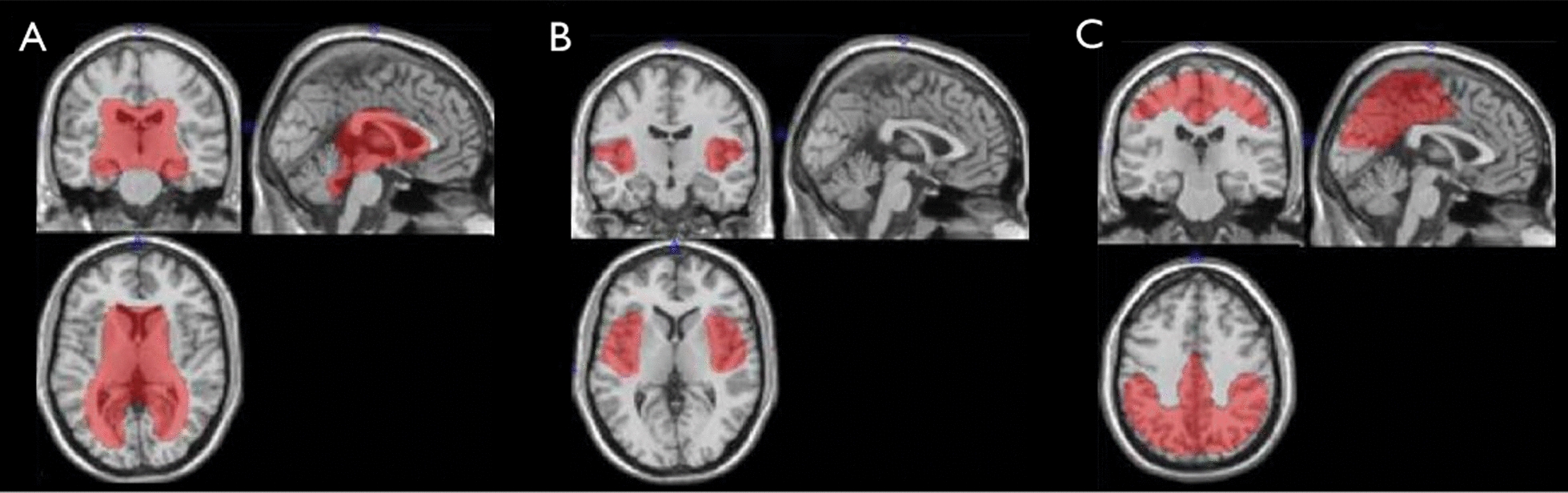
Methods: We measured CP volume, brain parenchyma, and cerebrospinal fluid (CSF) spaces associated with disproportionately enlarged subarachnoid space hydrocephalus (DESH), an imaging feature of normal-pressure hydrocephalus, in community-dwelling older adults aged ≥ 65 years without dementia.
Results: In 1,370 participants, lower Mini-Mental State Examination (MMSE) scores were significantly associated with higher CP volume, even after adjusting for DESH-related CSF space and brain parenchymal volume. CP volume was more strongly associated with MMSE scores than DESH-related CSF space and brain parenchymal volume. History of smoking, white matter hyperintensity, enlarged perivascular spaces, age, body mass index, and diabetes mellitus were also associated with increased CP volume.
Conclusions: CP volume may be a highly sensitive imaging marker of cognitive decline in community-dwelling older adults without dementia, as it is linked to cognitive decline independently of brain parenchyma and CSF volumes. Our findings emphasize the importance of investigating CP volume increase to maintain cognitive function in older individuals. Accordingly, further longitudinal studies are required.
Keywords: Cerebrospinal fluid; Choroid plexus; Cognitive impairment.
© 2024. The Author(s).
Conflict of interest statement
Declarations. Conflict of interest: The authors declare no competing interests. Ethics approval and consent to participate: The Research Ethics Committee of Kumamoto University (Kumamoto, Japan [approval number: GENOME-333]) approved this study. All participants provided written informed consent prior to data collection in accordance with the Declaration of Helsinki. Consent for publication: Not applicable.
Figures
References
MeSH terms
Grants and funding
- JP23dk0207053/the Japan Agency for Medical Research and Development
- JP23dk0207053/the Japan Agency for Medical Research and Development
- JP23dk0207053/the Japan Agency for Medical Research and Development
- JP23dk0207053/the Japan Agency for Medical Research and Development
- 23K14803/the Japan Society for the Promotion of Science
LinkOut - more resources
Full Text Sources
Medical
Miscellaneous
