. 2024 Dec 18;29(1):593.
doi: 10.1186/s40001-024-02181-z.

Mitochondria-targeted antioxidant mitoquinone mitigates vitrification-induced damage in mouse ovarian tissue by maintaining mitochondrial homeostasis via the p38 MAPK pathway

Affiliations

Mitochondria-targeted antioxidant mitoquinone mitigates vitrification-induced damage in mouse ovarian tissue by maintaining mitochondrial homeostasis via the p38 MAPK pathway

Tianqi Du et al. Eur J Med Res. .

Abstract

Objective: Ovarian tissue cryopreservation has become a promising alternative for fertility preservation in cancer patients, allowing ovarian tissue to be stored for future autotransplantation. Oxidative stress damage occurring during the cryopreservation process may impact tissue quality and function. This study aims to investigate the protective effects and potential mechanisms of Mitoquinone (MitoQ), a mitochondria-targeted derivative of the antioxidant ubiquinone, during the vitrification of ovarian tissue in mice.

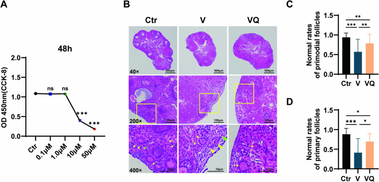
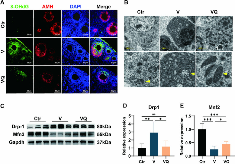
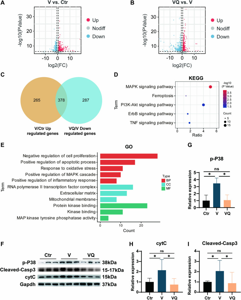
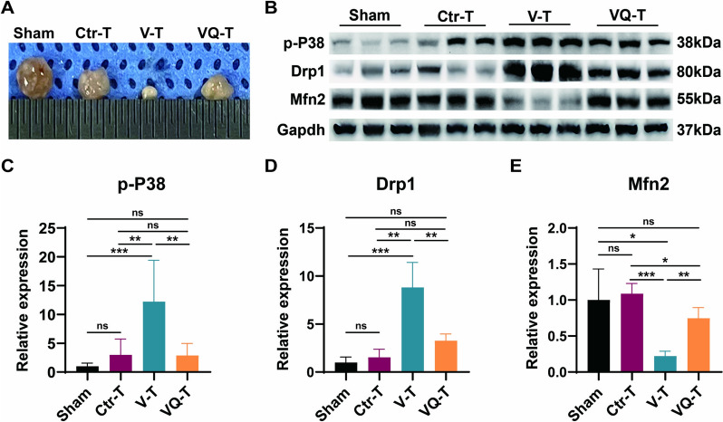
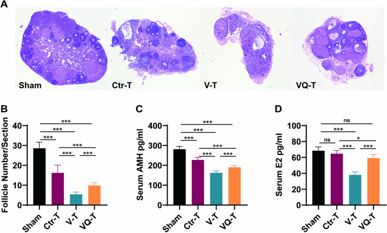
Methods: KGN cells were treated with various concentrations (0.1, 1, 10, and 50 μM) of MitoQ to determine the optimal concentration. Female ICR mice were divided into three groups: control, conventional vitrification, and MitoQ-supplemented vitrification. Ovarian samples were cryopreserved, thawed, and assessed for tissue morphology using Hematoxylin and Eosin (H&E) staining, and mitochondrial changes using immunofluorescence, transmission electron microscopy, and Western blot analysis. RNA sequencing (RNA-seq) was employed to explore potential protective mechanisms. Autotransplantation experiments were conducted, and the long-term effects of MitoQ on ovarian function were evaluated by counting follicle numbers through H&E staining and measuring serum estradiol and AMH levels using ELISA.

Results: MitoQ at 1 μM was found to be the optimal concentration for maintaining follicular morphology after vitrification. It effectively reduced mitochondrial oxidative damage, preserved mitochondrial morphology, and regulated the expression of mitochondrial dynamics proteins (Drp1 and Mfn2). RNA-seq and Western blot analyses revealed that MitoQ inhibited the p38 MAPK pathway, thereby reducing apoptosis. Additionally, autotransplantation experiments showed that MitoQ treatment significantly increased follicle counts, estradiol (E2), and anti-Müllerian hormone (AMH) levels compared to conventional vitrification.

Conclusions: MitoQ effectively mitigates vitrification-induced oxidative damage, maintains mitochondrial homeostasis, and preserves both follicular reserve and endocrine function. These findings suggest that MitoQ is a valuable adjunct in ovarian tissue cryopreservation and could significantly improve fertility preservation outcomes for cancer patients.

Keywords: Fertility preservation; Mitochondrial homeostasis; Mitoquinone; Ovarian tissue cryopreservation; Oxidative stress; p38 MAPK pathway.


Conflict of interest statement

Declarations. Ethics approval and consent to participate: This study was approved by the Ethics Committee of the Affiliated Suzhou Hospital of Nanjing Medical University, Suzhou Municipal Hospital (Approval No. K2023097). All procedures followed the guidelines of the institution’s Animal Care and Use Committee and were in accordance with international standards for the ethical treatment of animals. Competing interests: The authors declare no competing interests.

Figures

Fig. 1
Fig. 1
MitoQ enhances the proportion of morphologically normal primordial follicles and reduces DNA oxidative damage post-vitrification. A KGN cells were treated with varying concentrations of MitoQ (0.1 μM, 1.0 μM, 10 μM, and 50 μM) for 48 h. Cell viability was assessed using the CCK-8 assay, and absorbance was measured at 450 nm. Data are presented as mean ± SD (n = 4). B Representative images of ovarian tissues stained with H&E staining from each group under different magnifications (×4, ×20, ×40). Morphologically normal primordial follicles are indicated by green arrows, and morphologically normal primary follicles by yellow arrows. Morphologically abnormal primordial follicles are indicated by green triangles, and morphologically abnormal primary follicles by yellow triangles. Scale bars: 500 μm (×4), 100 μm (×20), and 10 μm (×40). C Statistical analysis of the proportion of morphologically normal primordial follicles in each group (n = 4). Data are expressed as mean ± SD. Statistical significance: *P < 0.05, ***P < 0.001. D Statistical analysis of the proportion of morphologically normal primary follicles in each group (n = 4). Data are expressed as mean ± SD. Statistical significance: *P < 0.05, ***P < 0.001
Fig. 2
Fig. 2
MitoQ improves mitochondrial morphology and homeostasis post-vitrification. A Immunofluorescence staining of ovarian tissues showing the localization of 8-hydroxy-2′-deoxyguanosine (8-OHdG) and anti-Müllerian hormone (AMH), with DAPI co-staining to assess DNA oxidative damage in ovarian tissues from each group (×20 magnification, n = 4). Representative images demonstrate that oxidative damage predominantly occurs in the granulosa cell mitochondria. B Transmission electron microscopy (TEM) images of mitochondrial morphology in ovarian tissues from each group (×4200 magnification, n = 3). Arrows indicate mitochondria. CE Western blot analysis of mitochondrial dynamics-related proteins Drp1 and Mfn2 in ovarian tissues from each group, with GAPDH as the loading control. Data are presented as mean ± SD (n = 3, **P < 0.01, ***P < 0.001). Ctr fresh control group, V conventional vitrification group, VQ MitoQ-supplemented vitrification group
Fig. 3
Fig. 3
Effects of vitrification and MitoQ-supplemented vitrification on gene expression and validation in mice ovaries. A Volcano plot showing differentially expressed genes (DEGs) between the V group and Ctr group. Fold change (FC) represents the ratio of transcripts per million (TPM), and the P-value is calculated by DESeq2 using a negative binomial distribution. Red and blue dots represent upregulated and downregulated genes, respectively, with |FC| > 1.5 and P < 0.05. B Volcano plot showing DEGs between the VQ group and V group. FC represents the fold change in TPM, and the P-value is calculated by DESeq2. Red and blue dots represent upregulated and downregulated genes, respectively, with |FC| > 1.5 and P < 0.05. C Venn diagram showing the overlap of upregulated DEGs in the V vs. Ctr group and downregulated DEGs in the VQ vs. V group, identifying 378 intersecting genes. D KEGG pathway enrichment analysis of the 378 genes significantly upregulated in the V group. The x-axis represents the ratio of enriched genes to the total number of background genes in the pathway, and the y-axis represents the enriched pathways. E GO enrichment analysis was conducted using the DAVID online tool, identifying the top 20 pathways for the 378 significantly upregulated genes in the V group. These pathways indicate relevant biological processes, cellular components, and molecular functions associated with these upregulated genes. FI Western blot analysis of p-P38, CytC, and Cleaved-Caspase3 protein levels in ovarian tissues from each group, with GAPDH as a loading control. Data are presented as mean ± SD (n = 3, *P < 0.05, **P < 0.01, ns: not significant). Ctr fresh control group, V conventional vitrification group, VQ MitoQ-supplemented vitrification group
Fig. 4
Fig. 4
Autotransplantation confirmed partial inhibition of the p38 MAPK pathway by adding MitoQ during vitrification-thawing. A Representative images of ovarian tissues from each group 4 weeks after transplantation. BE Western blot analysis of p-P38, Drp1, and Mfn2 protein levels in ovarian tissues from each group, with GAPDH as a loading control. Data are presented as mean ± SD (n = 3, *P < 0.05, **P < 0.01, ***P < 0.001, ns: not significant). Ctr fresh control group, V conventional vitrification group, VQ MitoQ-supplemented vitrification group
Fig. 5
Fig. 5
Autotransplantation confirmed the restoration of ovarian reserve and endocrine function by MitoQ during vitrification-thawing. A H&E staining of the largest cross-sections of ovarian tissues from each group (n = 4). B Statistical analysis of follicle counts in ovarian tissues from each group (n = 5, ***P < 0.001). C, D Statistical analysis of serum AMH and E2 levels in each group (n = 6, *P < 0.05, **P < 0.01, ***P < 0.001)

Similar articles

Cited by

References

    1. Xia C, Dong X, Li H, Cao M, Sun D, He S, et al. Cancer statistics in China and United States, 2022: profiles, trends, and determinants. Chin Med J. 2022;135(5):584–90. - PMC - PubMed
    1. Siegel RL, Giaquinto AN, Jemal A. Cancer statistics, 2024. CA Cancer J Clin. 2024;74(1):12–49. - PubMed
    1. Meirow D, Biederman H, Anderson RA, Wallace WH. Toxicity of chemotherapy and radiation on female reproduction. Clin Obstet Gynecol. 2010;53(4):727–39. - PubMed
    1. Stroud JS, Mutch D, Rader J, Powell M, Thaker PH, Grigsby PW. Effects of cancer treatment on ovarian function. Fertil Steril. 2009;92(2):417–27. - PubMed
    1. Spath MA, Braat DDM. Iatrogenic and non-iatrogenic causes of female fertility loss that may indicate fertility preservation. Acta Obstet Gynecol Scand. 2019;98(5):559–62. - PubMed

MeSH terms

LinkOut - more resources