. 2024 Dec 18;22(1):605.
doi: 10.1186/s12964-024-01935-w.

PD-1 interactome in osteosarcoma: identification of a novel PD-1/AXL interaction conserved between humans and dogs

Affiliations

PD-1 interactome in osteosarcoma: identification of a novel PD-1/AXL interaction conserved between humans and dogs

Katarzyna Dziubek et al. Cell Commun Signal. .

Abstract

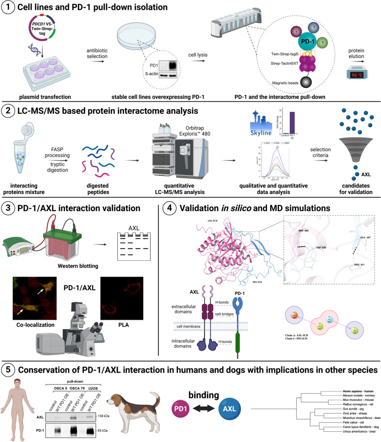
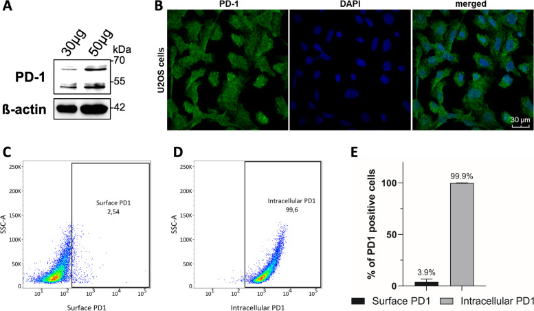
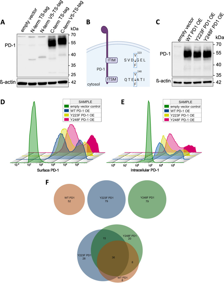
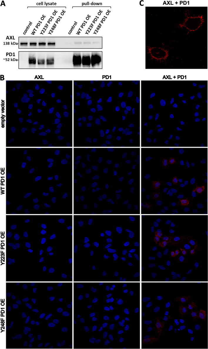
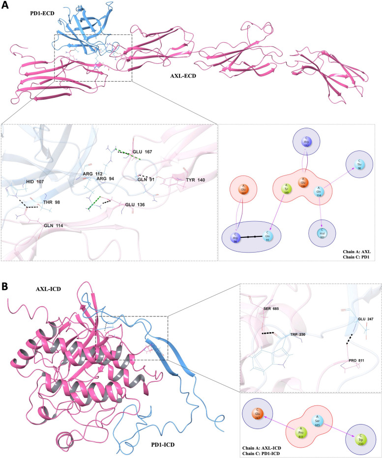
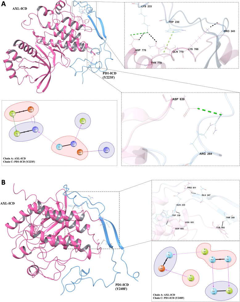
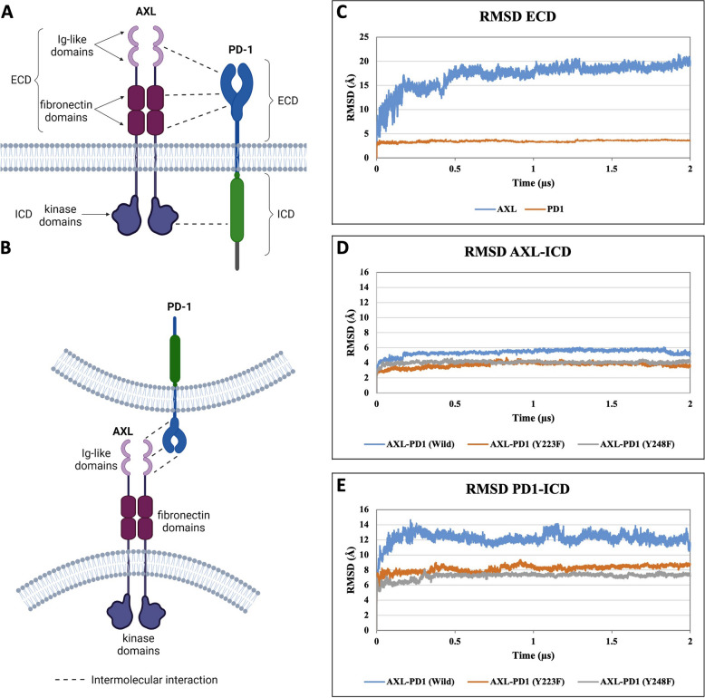
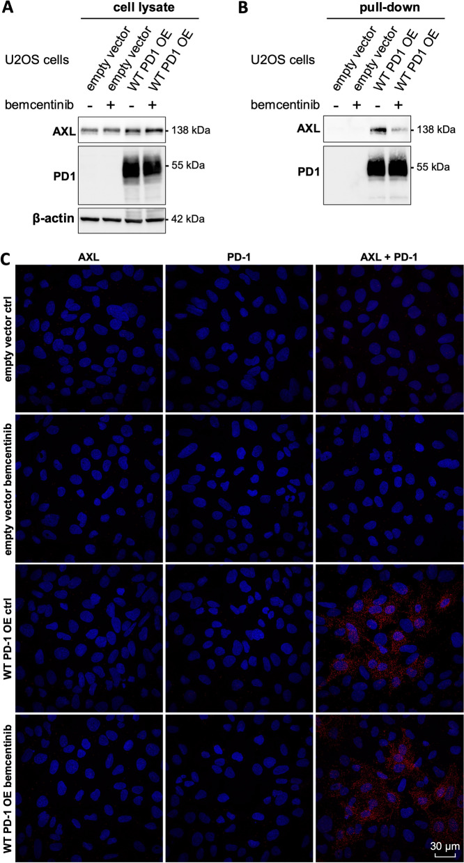
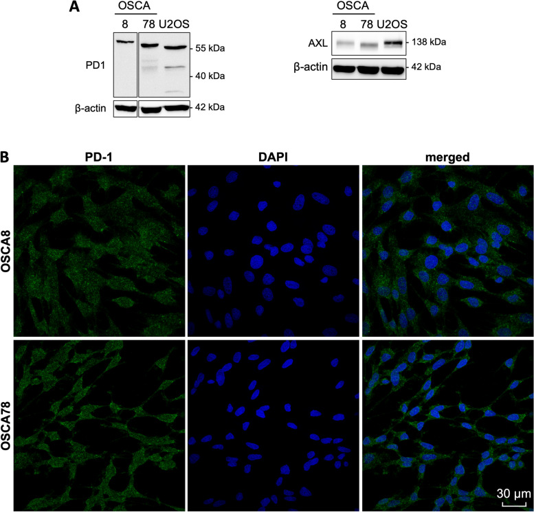
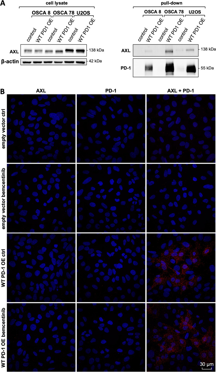
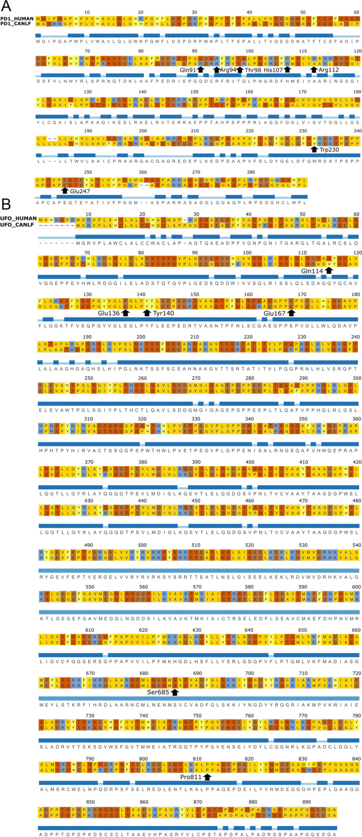
The PD-1/PDL-1 immune checkpoint inhibitors revolutionized cancer treatment, yet osteosarcoma remains a therapeutic challenge. In some types of cancer, PD-1 receptor is not solely expressed by immune cells but also by cancer cells, acting either as a tumor suppressor or promoter. While well-characterized in immune cells, little is known about the role and interactome of the PD-1 pathway in cancer. We investigated PD-1 expression in human osteosarcoma cells and studied PD-1 protein-protein interactions in cancer. Using U2OS cells as a model, we confirmed PD-1 expression by western blotting and characterized its intracellular as well as surface localization through flow cytometry and immunofluorescence. High-throughput analysis of PD-1 interacting proteins was performed using a pull-down assay and quantitative mass spectrometry proteomic analysis. For validation and molecular modeling, we selected tyrosine kinase receptor AXL-a recently reported cancer therapeutic target. We confirmed the PD-1/AXL interaction by immunoblotting and proximity ligation assay (PLA). Molecular dynamics (MD) simulations uncovered binding affinities and domain-specific interactions between extracellular (ECD) and intracellular (ICD) domains of PD-1 and AXL. ECD complexes exhibited strong binding affinity, further increasing for the ICD complexes, emphasizing the role of ICDs in the interaction. PD-1 phosphorylation mutant variants (Y223F and Y248F) did not disrupt the interaction but displayed varying strengths and binding affinities. Using bemcentinib, a selective AXL inhibitor, we observed reduced binding affinity in the PD-1/AXL interaction, although it was not abrogated. To facilitate the future translation of this finding into clinical application, we sought to validate the interaction in canine osteosarcoma. Osteosarcoma spontaneously occurs at significantly higher frequency in dogs and shares close genetic and pathological similarities with humans. We confirmed endogenous expression of PD-1 and AXL in canine osteosarcoma cells, with PD-1/AXL interaction preserved in the dog cells. Also, the interacting residues remain conserved in both species, indicating an important biological function of the interaction. Our study shed light on the molecular basis of the PD-1/AXL interaction with the implication for its conservation across species, providing a foundation for future research aimed at improving immunotherapy strategies and developing novel therapeutic approaches.

Keywords: AXL; Cancer-intrinsic PD-1; Comparative medicine; Immune checkpoints; Osteosarcoma; PD-1; Protein conservation.


Conflict of interest statement

Declarations. Competing interests: The authors declare no competing interests.

Figures

Fig. 1
Fig. 1
The experimental pipeline used to study the PD-1 protein–protein interactions and validation of PD-1 binding to AXL. Created with BioRender.com
Fig. 2
Fig. 2
PD-1 is endogenously expressed by U2OS human osteosarcoma cells (A) western blot analysis revealed the presence of PD-1 in U2OS cells, detected as two bands around 55 kDa and 70 kDa. The membrane was treated with stripping buffer and restained for ß-actin used as a loading control; (B) immunofluorescence analysis confirmed PD-1 expression in U2OS osteosarcoma cells, the staining was performed with Alexa Fluor™ 488 Tyramide SuperBoost™ and images were acquired with Olympus Fluoview FV3000 confocal microscope, using 60X objective with oil immersion. Nuclei were stained with DAPI; flow cytometry analysis revealed spontaneous expression of both (C) surface and (D) intracellular PD-1 in nearly all U2OS cells (more than 99%). The dot plots show data from a representative experiment; (E) demonstrates the mean percentage of the surface and intracellular expression of PD-1 calculated from four independent flow cytometry experiments. Error bars display standard deviation (SD)
Fig. 3
Fig. 3
Implementation of PD-1 overexpression allows for high throughput, LC–MS/MS based studies of the PD-1 interactome and the impact of PD-1 mutations on protein binding. (A) western blot analysis demonstrating the difference in PD-1 overexpression pattern in U2OS cells upon the N-terminal and C-terminal tagging of PD-1 protein; (B) schematic overview of the mutations introduced in PD-1 protein. (C) Stable expression of PD-1 in U2OS cells confirmed by western blot; (D) when overexpressed in U2OS cells, PD-1 protein is expressed on the surface and (E) intracellularly. The surface and intracellular staining performed for flow cytometry analysis revealed that upon overexpression PD-1 is expressed both extra- and intracellularly. Flow cytometry data were analyzed with FlowJo v10.8.1 flow cytometry analysis software (BD Biosciences); (F) DeepVenn diagram illustrating the number of proteins identified by LC–MS/MS analysis from the pull-down experiment, which are potential protein candidates for the interaction with PD-1 in WT, Y223F and Y248F overexpressing U2OS cells; the diagram illustrates distribution of shared and unique proteins identified in the WT and mutant PD-1 overexpressing samples
Fig. 4
Fig. 4
Pull-down immunoblotting and proximity ligation assay confirm the interaction between PD-1 and AXL, which is not disrupted by the mutations in PD-1 phosphorylation sites. (A) western blotting of the pull-down samples confirms the interaction between PD-1 and AXL. Stripping buffer was used to remove AXL staining from the membrane, which was further reprobed for PD-1 used both as a loading control and positive control for the pull-down experiment; (B) PLA was performed between PD-1 and AXL, samples were mounted with mounting media with DAPI. The figure demonstrates representative Z-stack images of the PLA (red). Specimens were visualized by confocal microscopy with 60X oil immersion lens, bar = 30 µm; (C) A representative image of a single focal plane acquired at 120X magnification indicating that the interaction between PD-1 and AXL in WT PD-1 OE U2OS cells localizes to the cellular membrane
Fig. 5
Fig. 5
3D interaction diagram of docked AXL-PD-1 domain complexes (A) AXL-ECD and PD-1-ECD, (B) AXL-ICD and PD-1-ICD (WT). Interacting residues (line representation) are depicted in zoomed out image. Interactions shown in dashed line with following color scheme as applicable: H-bond (black), salt-bridge (green), pi-pi stacking (lime), and pi-cation (dark green)
Fig. 6
Fig. 6
3D interaction diagram of docked AXL-PD-1 domain complexes (A) AXL-ICD and PD-1-ICD (Y223F) and (B) AXL-ICD and PD-1-ICD (Y248F) complex. Interacting residues (line representation) are depicted in zoomed out image. Interactions shown in dashed line with following color scheme as applicable: H-bond (black), salt-bridge (green), pi-pi stacking (lime), and pi-cation (dark green)
Fig. 7
Fig. 7
Root mean square deviation (RMSD) analysis throughout the molecular dynamics simulation trajectories; (A) schematic illustration of possible intermolecular interactions between AXL and PD-1, when both AXL and PD-1 are embedded within the same cell membrane; (B) when AXL and PD-1 are present in different cell membranes; molecular dynamics simulation trajectories of (C) AXL and PD-1 ECDs, (D) AXL ICD when interacting with PD-1 ICDs (WT and mutants), (E) PD-1 ICDs (WT and mutants) when complexed with AXL ICD. A and B created with BioRender.com
Fig. 8
Fig. 8
Western blotting of PD-1 pull-down demonstrates that bemcentinib treatment alters the PD-1/AXL interaction in U2OS cells but does not abrogate it as evidenced by the proximity ligation assay; (A) immunoblotting of whole cell lysate used for PD-1 pull-down. Samples were collected after 24 h treatment with 2.5 μM bemcentinib or equivalent volume of DMSO, which was used for bemcentinib reconstitution; (B) western blotting of samples after PD-1 pull-down demonstrated a decreased level of AXL upon bemcentinib treatment, indicating lower affinity of PD-1/AXL interaction upon AXL inhibition. The membranes were reprobed for PD-1 and β-actin used as a positive control for the pull-down experiment and loading control, respectively; (C) PLA performed between PD-1 and AXL shows no visible changes in the PD-1/AXL interaction upon treatment with bemcentinib. Nuclear staining was performed with DAPI, samples were mounted with mounting media. Specimens were visualized by confocal microscopy with 60X oil immersion lens, bar = 30 µm
Fig. 9
Fig. 9
PD-1 and AXL are endogenously expressed by canine osteosarcoma cell lines as indicated by western blotting and immunofluorescence. Western blot analysis demonstrated that (A) PD-1 and AXL are naturally expressed in canine osteosarcoma cell lines OSCA8 and OSCA78. Human osteosarcoma U2OS cells were used as a control for antibody specificity; (B) canine PD-1 expression was additionally confirmed by immunofluorescence analysis performed with Alexa Fluor™ 488 Tyramide SuperBoost™. Images were acquired with confocal microscope, using 60X objective with oil immersion. Nuclei were stained with DAPI
Fig. 10
Fig. 10
Immunoblotting of canine PD-1 pull-down and proximity ligation assay confirm that the PD-1/AXL interaction occurs in canine cells; (A) western blotting of the pull-down samples confirming the interaction between PD-1 and AXL. A stripping buffer was used to remove AXL staining from the membrane, which was further reprobed either for β-actin or PD-1 as a loading control or positive control for the pull-down experiment, respectively; (B) PLA performed between canine PD-1 and AXL, nuclear staining was performed with DAPI. The figure demonstrates representative Z-stack images of the PLA (red). Specimens were visualized by confocal microscopy with 60X oil immersion lens, bar = 30 µm
Fig. 11
Fig. 11
Sequence alignment of canine and reference human PD-1 and AXL with highlighted amino acids involved in PD-1/AXL interaction interface in human. Figure suggests a conservation of (A) PD-1 and (B) AXL amino acids involved in PD-1/AXL interaction interface with minor dissimilarity observed. Alignment suggests that the interaction might be present also in dog, highlighting further implications of these findings over multiple species shown in Supplementary Fig. 6 A, B and C. Figure created with EMBL-EBI Job Dispatcher [63]

Similar articles

References

    1. Bardhan, K., Anagnostou, T., & Boussiotis, V. A. (2016). The PD1: PD-L1/2 pathway from discovery to clinical implementation. Front Immunol, 7(DEC). 10.3389/fimmu.2016.00550. - PMC - PubMed
    1. Topalian SL, Drake CG, Pardoll DM. Targeting the PD-1/B7-H1(PD-L1) pathway to activate anti-tumor immunity. Curr Opin Immunol. 2012;24(2):207–12. 10.1016/j.coi.2011.12.009. - PMC - PubMed
    1. Cole S, Gianferante DM, Zhu B, Mirabello L. Osteosarcoma: A Surveillance, Epidemiology, and End Results program-based analysis from 1975 to 2017. Cancer. 2022;128(11):2107–18. 10.1002/CNCR.34163. - PMC - PubMed
    1. Mirabello L, Troisi RJ, Savage SA. International osteosarcoma incidence patterns in children and adolescents, middle ages, and elderly persons. Int J Cancer: J Int Du Cancer. 2009;125(1):229. 10.1002/IJC.24320. - PMC - PubMed
    1. Rastogi, S., Aggarwal, A., Tiwari, A., & Sharma, V. Chemotherapy in nonmetastatic osteosarcoma: Recent advances and implications for developing countries. Journal of Glob Oncol, 2018(4). 10.1200/JGO.2016.007336. - PMC - PubMed

MeSH terms

Substances

LinkOut - more resources