Review
. 2024 Dec 19;22(1):767.
doi: 10.1186/s12951-024-03037-8.

Regulation of dynamic spatiotemporal inflammation by nanomaterials in spinal cord injury

Affiliations
Review

Regulation of dynamic spatiotemporal inflammation by nanomaterials in spinal cord injury

Zeping Liu et al. J Nanobiotechnology. .

Abstract

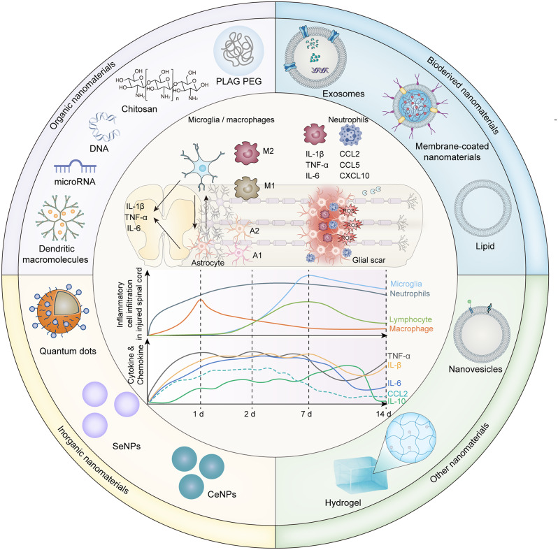
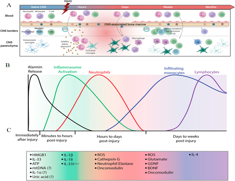
Spinal cord injury (SCI) is a common clinical condition of the central nervous system that can lead to sensory and motor impairment below the injury level or permanent loss of function in severe cases. Dynamic spatiotemporal neuroinflammation is vital to neurological recovery, which is collectively constituted by the dynamic changes in a series of inflammatory cells, including microglia, neutrophils, and astrocytes, among others. Immunomodulatory nanomaterials can readily improve the therapeutic effects and simultaneously overcome various drawbacks associated with treatment, such as the off-target side effects and loss of bioactivity of immune agents during circulation. In this review, we discuss the role of dynamic spatiotemporal inflammation in secondary injuries after SCI, elaborate on the mechanism of action and effect of existing nanomaterials in treating SCI, and summarize the mechanism(s) whereby they regulate inflammation. Finally, the challenges and prospects associated with using nanotechnology to modulate immunotherapy are discussed to provide new insights for future treatment. Deciphering the intricate spatiotemporal mechanisms of neuroinflammation in SCI requires further in-depth studies. Therefore, SCI continues to represent a formidable challenge.

Keywords: Astrocyte; Inflammation; Microglia; Nanomaterials; Spatiotemporal dynamic; Spinal cord injury.


Conflict of interest statement

Declarations. Ethics approval and consent to participate: Not applicable. Consent for publication: Not applicable. Competing interests: The authors declare no competing interests.

Figures

Scheme 1
Scheme 1
A schematic depiction of the inflammatory responses mediated by immune cells and cytokines, their dynamic shifts in the context of traumatic spinal cord injury, and the classification and structure of nanomaterials used for inflammation modulation
Fig. 1
Fig. 1
Temporal transcriptomic changes post-spinal cord injury (SCI), as revealed by population-based (bulk-RNA-seq) and single-cell RNA sequencing (scRNA-seq): A) Schematic representation of the tissue sampling process for bulk-RNA-seq and scRNA-seq. B) The t-distributed Stochastic Neighbor Embedding (t-SNE) plot demonstrates consistent sequencing outcomes across different samples. C) Hierarchical clustering of 39 samples showing the expression heatmap of each module across all samples. D) Gene ontology (GO) terms for seven gene modules and their average gene expression changes over time post-SCI. E) Pie chart illustrating the number of genes within each module. F) Overview of the 10× Genomics scRNA-seq experimental workflow. G) Visualization of spinal cord cells from different samples using Uniform Manifold Approximation and Projection (UMAP). H) Line graph depicting the temporal changes in the relative proportions of 12 major cell types identified through scRNA-seq. Used under the terms of the Creative Commons Attribution 4.0 International License (CC BY 4.0). License details available at https://creativecommons.org/licenses/by/4.0/
Fig. 2
Fig. 2
Inflammation associated with central nervous system (CNS) injury. A) Compartmentalization of the immune response after CNS injury. B) Phases of molecular and cellular inflammation after CNS injury. C) Activity of specific inflammatory molecules at different time points corresponding to the injury. Reproduced from with permission [54, 55]. Licensed under CC BY 4.0. Copyright © 2023 Elsevier
Fig. 3
Fig. 3
After spinal cord injury, microglia extensively proliferate and accumulate at the injury boundary. AF) Confocal microscopic images displaying representative spinal cord sections from uninjured to 1–35 dpi. GP) Number of microglia in each group and the expression of apoptosis and proliferation markers. Reproduced with permission [72]. Licensed under CC BY 4.0
Fig. 4
Fig. 4
Therapeutic outcomes of RA@BSA@Cur NPs. A) Immunofluorescence and quantification of RAW264.7 cells cultured with different BSA-related nanoparticles (NPs) in the presence of lipopolysaccharide (LPS); B) Flow cytometry analysis and quantification of RAW264.7 cells cultured with various BSA-related NPs in the presence of LPS. The inhibition of the M1 phenotype and promotion of the M2 phenotype regulated by RA@BSA@Cur NPs may involve the NF-κB pathway. C) Changes in the expression of CD86 and CD206 under normal conditions and in the context of SCI. D) Ratio of CD80 + to CD206 + cells in different groups. Reproduced with permission [120]. Licensed under CC BY 4.0
Fig. 5
Fig. 5
Therapeutic outcomes of ET@PPP-ACPP. A) Preparation of PPP and ET@PPP-ACPP. B) Characterization of ET@PPP-ACPP. C) Spinal cord injury (SCI)-targeting effect of PPP-ACPP NPs. D) Proinflammatory cytokine levels in the spinal cords of SCI mice, analyzed using the Luminex analysis system 24 h post-SCI. E) Levels of proinflammatory and anti-inflammatory cytokines in the spinal cord post-treatment. Reproduced with permission [115]. Copyright © 2023 Wiley
Fig. 6
Fig. 6
Therapeutic outcomes of NPs-pOXR1-Lip. A) Characterization of NPs-pOXR1-Lip. B-D) NPs-pOXR1-Lip decreases apoptosis following spinal cord injury. (SCI). EG) NPs-pOXR1-Lip diminishes fibrotic scar tissue by influencing the inflammatory response. H, I) NPs-pOXR1-Lip facilitates functional recovery post-SCI. Reproduced from with permission [137]. Licensed under CC BY 4.0
Fig. 7
Fig. 7
Preparation and therapeutic effects of CCR2-targeted PLGA/curcumin nanoparticles (CCR2-MM@PLGA/Cur/NPs). A) Preparation of CCR2-MM@PLGA/Cur/NPs. B) Average diameters and zeta potentials. In vitro drug release characteristics of PLGA/Cur, MM@PLGA/Cur, and CCR2-MM@PLGA/Cur. Results from western blot analyses showing the expression levels of CCR2, TNFR2, TLR4, and CD36 in PLGA/NPs, MM@PLGA/NPs, CCR2-MM@PLGA/NPs, RAW 264.7, and RAW 264.7-CCR2 cell lines. C) Analysis via FCM of CCR2 and specific protein biomarkers (CD206, CD11c, and F4/80) expression in CCR2-MM@PLGA/Cur, CCR2-MM, and RAW 264.7-CCR2 cells. D) Immunostaining representative images showing CD86 (in red) and CD206 (in green) in BV2 cells across various experimental groups. E) Quantitative immunofluorescence of CD86 (indicating pro-inflammatory) and CD206 (indicating anti-inflammatory) in microglia, and the relative expression of pro-inflammatory genes (TNF-α, IL-1β, IL-6, and iNOS), normalized to control levels [140]. Reproduced with permission. Copyright © 2023 Wiley
Fig. 8
Fig. 8
Aux@BT nanoparticles alleviate spinal cord injury by generating hydrogen. A) Aux@BT nanoparticle synthesis and characterization; X = 1, 3, or 5. B) Principle underlying the generation of H2. C) Illustration of the ultrasound-assisted piezoelectric catalytic therapy for rats with spinal cord injury. Numbers of inflammatory cells and levels of IL-1γ and IL-6 in the blood of rats. D) Representative histological images corresponding to the expression of TNF-α, IL-1γ, and IL-6. Reproduced with permission [158]. Copyright © 2023 Wiley
Fig. 9
Fig. 9
Assembly and therapeutic effects of MP2-TK@ RU nanoparticles. A) Synthesis of the precursor MP2-TK. B) Self-assembly and ROS-responsive behavior of MP2-TK@RU NPs. C) Treatment principles of spinal cord injury. D) Representative immunofluorescence images of injured spinal cords eight weeks post-different treatments. E) Relative intensity of Iba-1 immunofluorescence in injured spinal cords. F, G) Magnified immunofluorescence images of TNF-α. H, I) Relative intensity of TNF-α H) and IL-6 I) immunofluorescence in injured spinal cords. Reproduced with permission [107]. Copyright © 2023 American Chemical Society

Similar articles

Cited by

References

    1. Tamburin S, Filippetti M, Mantovani E, Smania N, Picelli A. Spasticity following brain and spinal cord injury: assessment and treatment. Curr Opin Neurol. 2022;35(6):728–40. - PubMed
    1. Albashari AA, He Y, Luo Y, Duan X, Ali J, Li M, et al. Local spinal cord injury treatment using a dental pulp stem cell encapsulated H2S releasing multifunctional injectable hydrogel. Adv Healthc Mater. 2023;13(9):e2302286. - PMC - PubMed
    1. Alonso F, Hart DJ. Advanced imaging techniques in cervical spinal cord trauma. World Neurosurg. 2015;83(4):496–7. - PubMed
    1. Rao J-S, Zhao C, Wei R-H, Feng T, Bao S-S, Zhao W, et al. Neural regeneration therapy after spinal cord injury induces unique brain functional reorganizations in rhesus monkeys. Ann Med. 2022;54(1):1867–83. - PMC - PubMed
    1. Badhiwala JH, Wilson JR, Witiw CD, Harrop JS, Vaccaro AR, Aarabi B, et al. The influence of timing of surgical decompression for acute spinal cord injury: a pooled analysis of individual patient data. Lancet Neurol. 2021;20(2):117–26. - PubMed

LinkOut - more resources