. 2024 Dec 18;15(1):467.
doi: 10.1186/s13287-024-04109-0.

Transcriptome-aligned metabolic profiling by SERSome reflects biological changes following mesenchymal stem cells expansion

Affiliations

Transcriptome-aligned metabolic profiling by SERSome reflects biological changes following mesenchymal stem cells expansion

Xinyuan Bi et al. Stem Cell Res Ther. .

Abstract

Background: Mesenchymal stem cells (MSCs) are widely applied in the treatment of various clinical diseases and in the field of medical aesthetics. However, MSCs exhibit greater heterogeneity limited stability, and more complex molecular and mechanistic characteristics compared to conventional drugs, making rapid and precise monitoring more challenging.

Methods: Surface-enhanced Raman spectroscopy (SERS) is an ultrasensitive, tractable and low-cost fingerprinting technique capable of identifying a wide range of molecules related to biological processes. Here, we employed SERS for reproducible quantification of ultralow concentrations of molecules and utilized spectral sets, termed SERSomes, for robust and comprehensive intracellular multi-metabolite profiling.

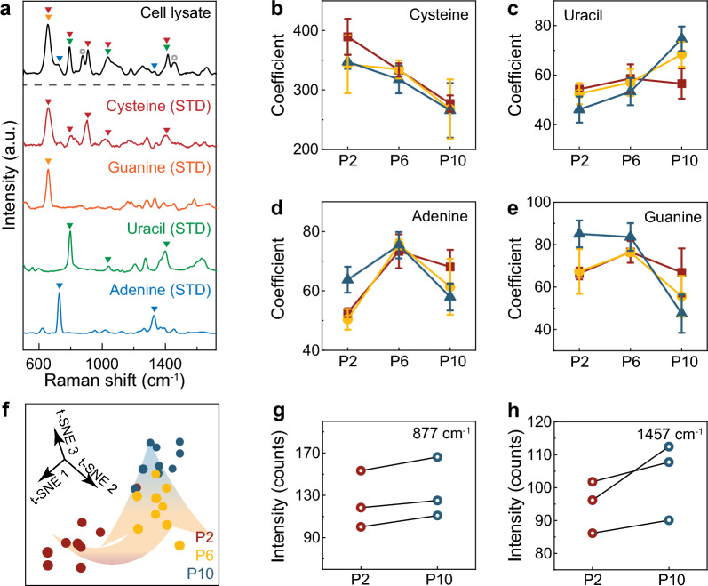
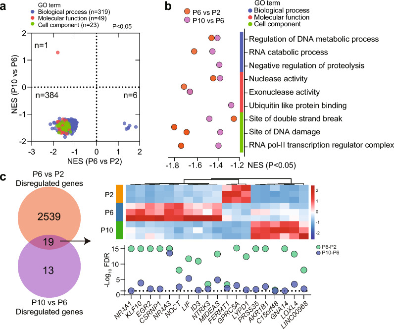
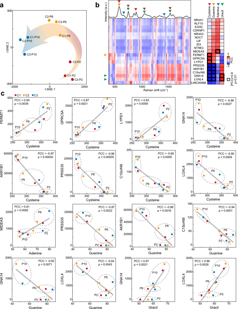
Results: We revealed that with increasing passage number, there is a gradual decline in cell expansion efficiency, accompanied by significant changes in intracellular amino acids, purines, and pyrimidines. By integrating these metabolic features detected by SERS with transcriptomic data, we established a correlation between SERS signals and biological changes, as well as differentially expressed genes.

Conclusion: In this study, we explore the application of SERS technique to provide robust metabolic characteristics of MSCs across different passages and donors. These results demonstrate the effectiveness of SERSome in reflecting biological characteristics. Due to its sensitivity, adaptability, low cost, and feasibility for miniaturized instrumentation throughout pretreatment, measurement, and analysis, the label-free SERSome technique is suitable for monitoring MSC expansion and offers significant advantages for large-scale MSC manufacturing.

Keywords: Mesenchymal stem cells (MSCs); Metabolism; Surface-enhanced Raman spectroscopy (SERS); Transcriptome.


Conflict of interest statement

Declarations. Ethics approval and consent to participate: This study was conducted at the School of Biomedical Engineering and Clinical Stem Cell Research Center, Shanghai Jiao Tong University. Approval was granted for a project titled “Cancer Immunotherapy Using Umbilical Cord Mesenchymal Stem Cells” in 2021 by the Renji Hospital Ethics Committee (No. KY2021-027). All participants provided informed consent. Consent for publication: Not applicable. Competing interests: The authors declare no competing interests.

Figures

Fig. 1
Fig. 1
Metabolic profiling of hUC-MSC lysates by SERS. a Workflow of hUC-MSC metabolome-transcriptomic analysis. b Bright-field images of hUC-MSCs (scale bar: 200 μm) and the SERS metabolic profiling of the corresponding cell lysates. For each lysate sample, a spectral set comprising 200 spectra were measured as displayed by the heatmaps. The mean spectra are provided for clarity. c The relative cell number, d The cell viability, e the passaging number, and f the expansion rate has been recorded along the 14-day culture. For each cell origin, the data are displayed by the mean values from three biological replicates. Error bar: standard deviation (n = 3). g Absorbance spectrum and h the hydrodynamic diameter of citrate-reduced Ag NPs. i Pearson’s correlation coefficients among the mean SERS spectra obtained from the C1-originated hUC-MSC lysates of different generations (P2, P6 and P10) and different biological replicates (S1, S2 and S3). j Pearson’s correlation coefficients among the mean SERS spectra obtained from the hUC-MSC lysates of different generations (P2, P6 and P10) and origins (C1, C2 and C3). k 2D t-SNE visualization of all the SERS spectra and l the highlighted t-SNE distribution of the SERS spectra obtained from the C1-originated hUC-MSC lysates. Source data are available in the Source Data file
Fig. 2
Fig. 2
Evaluation of SERS metabolic indicators for hUC-MSC expansion. a The mean SERS spectra of all hUC-MSC lysates (cell lysate) and the standard SERS spectra (STD) of pure metabolite solutions (cysteine, guanine, uracil and adenine). The characteristic peaks of the metabolites are exhibited by colored arrows. The coefficients of b cysteine, c uracil, d adenine and e guanine obtained by decomposing the lysate SERS spectra of C1 hUC-MSCs at different generations. The data are displayed by the mean value from three biological replicates (blue, yellow and red) with the error bar indicating the standard deviation (n = 3 technical replicates). f t-SNE visualization of the metabolic coefficients of C1 hUC-MSC lysates. For each cell generation, there are 9 datapoints obtained from three biological replicates, each being measured three times. The changes of SERS peak intensities at g 877 and h 1459 cm−1 along the expansion (from P2 to P10) of three replicates of C1 hUC-MSCs. The SERS bands of 877 and 1459 cm−1 are pointed out by black circles in panel a. Source data are available in the Source Data file
Fig. 3
Fig. 3
Transcriptomics analysis of MSCs. a Heat map of the altered mRNAs between P6 (n = 3) and P2 (n = 3) MSCs, as well as between P10 (n = 3) and P6 (n = 3) MSCs. b Volcano plot of gene alterations across different passages of MSCs. Significantly differential mRNAs (adjusted P. value < 0.05 and fold change > 1.5) are colored in red (up-regulated) and blue (down-regulated). c Significantly altered GO pathways. d Significantly altered KEGG pathways. e Relative mRNA levels of indicated genes. f Heat map of relative mRNAs levels of indicated genes. Data are represented as mean ± SEM. Source data are available in the Source Data file
Fig. 4
Fig. 4
Consistent alterations in cellular processes. a GSEA analysis of up-regulated and down-regulated pathways in P6 MSCs compared to P2 MSCs (x-axis) and in P10 MSCs compared to P6 (y-axis), based on GO subset in AmiGO database. Normalized enrichment scores (NES) of GO terms are plotted. b Quantitative analysis of altered pathways across different passages of MSCs based on GSEA analysis. (c) Venn diagram and heat map showing dis-regulated genes across different passages of MSCs. Source data are available in the Source Data file
Fig. 5
Fig. 5
Relation between SERS metabolic profiling and RNA expression. a t-SNE visualization of the 19 most significantly changed genes. b Pearson’s correlation coefficient between the RNA expression and the SERS spectral intensities (left) as well as the metabolite coefficients (right). The mean spectrum of all lysate SERS spectra is displayed with the characteristic peaks of the metabolites pointed by colored arrows for clarity. The correlation between RNA expressions and metabolite coefficients with |PCC|> 0.8 and p < 0.01 are exhibited by thick black boxes and c displayed in detail by scatter plots with the different cell generations labeled by dashed circles. Source data are available in the Source Data file

Similar articles

Cited by

  • Surface-Enhanced Raman Spectroscopy for Biomedical Applications: Recent Advances and Future Challenges.
    Lin LL, Alvarez-Puebla R, Liz-Marzán LM, Trau M, Wang J, Fabris L, Wang X, Liu G, Xu S, Han XX, Yang L, Shen A, Yang S, Xu Y, Li C, Huang J, Liu SC, Huang JA, Srivastava I, Li M, Tian L, Nguyen LBT, Bi X, Cialla-May D, Matousek P, Stone N, Carney RP, Ji W, Song W, Chen Z, Phang IY, Henriksen-Lacey M, Chen H, Wu Z, Guo H, Ma H, Ustinov G, Luo S, Mosca S, Gardner B, Long YT, Popp J, Ren B, Nie S, Zhao B, Ling XY, Ye J. Lin LL, et al. ACS Appl Mater Interfaces. 2025 Mar 19;17(11):16287-16379. doi: 10.1021/acsami.4c17502. Epub 2025 Feb 24. ACS Appl Mater Interfaces. 2025. PMID: 39991932 Free PMC article. Review.

References

    1. Ankrum JA, Ong JF, Karp JM. Mesenchymal stem cells: immune evasive, not immune privileged. Nat Biotechnol. 2014;32(3):252–60. - PMC - PubMed
    1. Caplan AI. Mesenchymal stem cells. J Orthop Res. 1991;9(5):641–50. - PubMed
    1. Chancellor D, et al. The state of cell and gene therapy in 2023. Mol Ther. 2023;31(12):3376–88. - PMC - PubMed
    1. Krampera M, Le Blanc K. Mesenchymal stromal cells: Putative microenvironmental modulators become cell therapy. Cell Stem Cell. 2021;28(10):1708–25. - PubMed
    1. Fung M, et al. Responsible translation of stem cell research: an assessment of clinical trial registration and publications. Stem Cell Rep. 2017;8(5):1190–201. - PMC - PubMed