. 2024 Dec 18;26(1):212.
doi: 10.1186/s13075-024-03424-4.

Role of TLR4 activation and signaling in bone remodeling, and afferent sprouting in serum transfer arthritis

Affiliations

Role of TLR4 activation and signaling in bone remodeling, and afferent sprouting in serum transfer arthritis

Gilson Goncalves Dos Santos et al. Arthritis Res Ther. .

Abstract

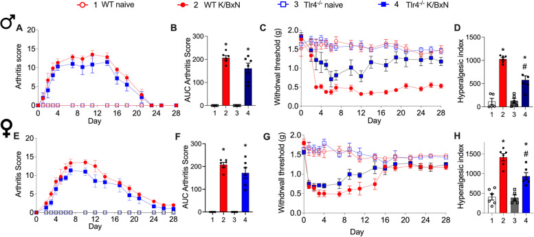
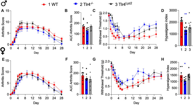
Background: In the murine K/BxN serum transfer rheumatoid arthritis (RA) model, tactile allodynia persists after resolution of inflammation in male and partially in female wild type (WT) mice, which is absent in Toll-like receptor (TLR)4 deficient animals. We assessed the role of TLR4 on allodynia, bone remodeling and afferent sprouting in this model of arthritis.

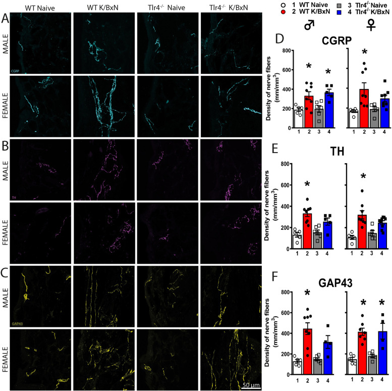
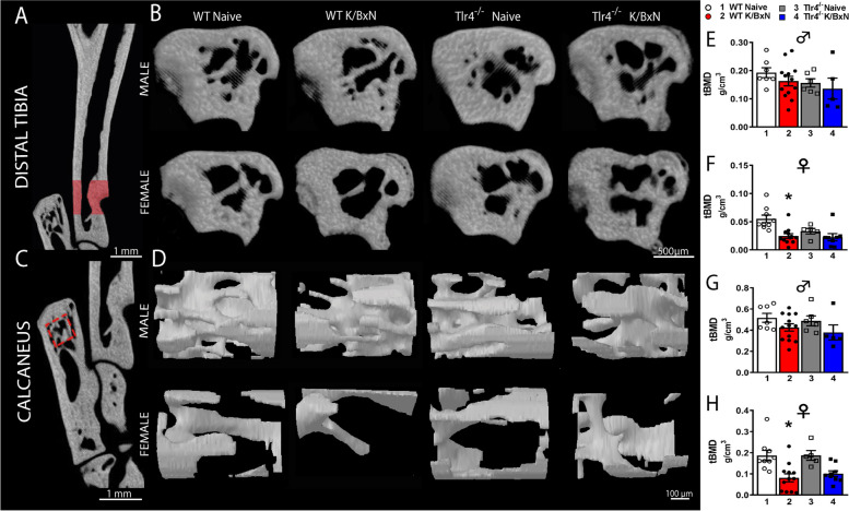
Methods: K/BxN sera were injected into male and female mice with conditional or stable TLR4 deletion and controls. Paw swelling was scored and allodynia assessed by von Frey filaments. At day 28, synovial neural fibers were visualized with confocal microscopy and bone density assayed with microCT. Microglial activity and TLR4 dimerization in spinal cords were examined by immunofluorescence and flow cytometry.

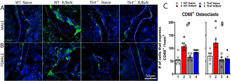
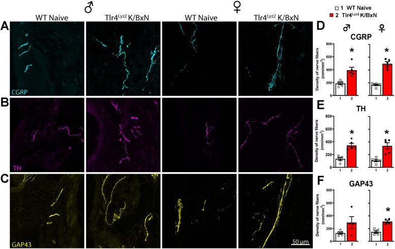
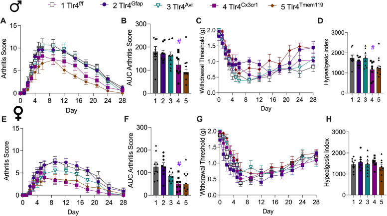
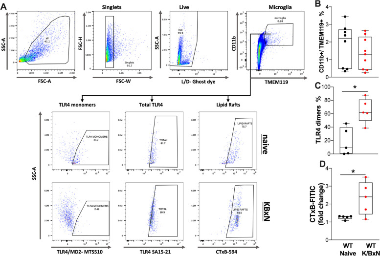
Results: In the synovium, K/BxN injected WT male and female mice showed robust increases in calcitonin gene related-peptide (CGRP+), tyrosine hydroxylase (TH)+ and GAP43+ nerve fibers. Trabecular bone density by microCT was significantly decreased in K/BxN WT female but not in WT male mice. The number of osteoclasts increased in both sexes of WT mice, but not in Tlr4-/- K/BxN mice. We used conditional strains with Cre drivers for monocytes/osteoclasts (lysozyme M), microglia (Tmem119 and Cx3CR1), astrocytes (GFAP) and sensory neurons (advillin) for Tlr4f/f disruption. All strains developed similar arthritis scores after K/BxN serum injection with the exception being the Tlr4Tmem119 mice which showed a reduction. Both sexes of Tlr4Lyz2, Tlr4Tmem119 and Tlr4Cx3cr1 mice displayed a partial reversal of the chronic pain phenotype but not in Tlr4Avil, and Tlr4Gfap mice. WT K/BxN male mice showed increases in spinal Iba1, but not GFAP, compared to Tlr4-/- male mice. To determine whether spinal TLR4 was indeed activated in the K/BxN mice, flow cytometry of lumbar spinal cords of WT K/BxN male mice was performed and revealed that TLR4 in microglia cells (CD11b+ /TMEM119+) demonstrated dimerization (e.g. activation) and a characteristic increase in lipid rafts.

Conclusion: These results demonstrated a complex chronic allodynia phenotype associated with TLR4 in microglia and monocytic cell lineages, and a parallel spinal TLR4 activation. However, TLR4 is dispensable for the development of peripheral nerve sprouting in this model.

Keywords: Arthritis; CGRP; Inflammation; Microglia; Pain; TLR4.


Conflict of interest statement

Declarations. Ethics approval and consent to participate: Animal experiments were approved by the UCSD Institutional Animal Care and Use Committee (IACUC). No human subjects or human material were included in these studies. Consent for publication: Not applicable. Competing interests: TLY and YIM are inventors listed in patent applications related to the topic of this paper and scientific co-founders of Raft Pharmaceuticals LLC. The terms of this arrangement have been reviewed and approved by the University of California San Diego, in accordance with its conflict-of-interest policies. Other authors declare that they have no competing interests.

Figures

Fig. 1
Fig. 1
Differential effects of TLR4 on clinical score and allodynia with K/BxN serum transfer. Male (A-D) and female (EH) WT mice and Tlr4−/− mice (were injected on days 0 and 2 with K/BxN sera and developed transiently increased clinical scores with paw inflammation (A, E) and allodynia (C, G). The AUCs of aggregate arthritis scores were calculated for all groups. All strains and sexes increased AUCs of arthritis scores compared to their respective naïve controls (B and F, *p = 0.0001 and one-way ANOVA with Tukey post hoc test). Associated allodynia resolved in the Tlr4−/− male mice but not in the WT male mice (C). The AUCs of aggregate hyperalgesic indices were calculated for all groups. Increased hyperalgesic indices were observed in male and female WT K/BxN mice compared to WT naïve (D and H, respectively, *p = 0.001 and *p = 0.0001, one-way ANOVA with Tukey post hoc test) and hyperalgesic indices for Tlr4−/− K/BxN mice compared to WT K/BxN mice, #p = 0.007 and p = 0.002). Data are represented as mean ± SEM with n = 6 per group
Fig. 2
Fig. 2
Arthritic mice have increased density of CGRP + , TH + and GAP43 + nerve fibers in the ankle joint. Representative confocal images of CGRP (A, cyan), TH (B, purple) and GAP43 (C, yellow) in ankle-joint sections (20 μm-thick) from naïve and serum injected WT and Tlr4−/− male and female mice. Significantly greater densities of CGRP+ (D), TH+ (E), and GAP43+ (F) nerve fibers were quantitated in WT serum injected compared to naïve males (p = 0.46; p = 0.003; p = 0.005 respectively) and females (p = 0.007, p = 0.0003, p = 0.0005 respectively). Male Tlr4−/− serum injected mice had a significant increase in the density of CGRP+ (D; p = 0.03), but not TH+ (E; p = 0.10) fibers and GAP43+ (F; p = 0.14) fibers, compared to naïve controls. Female Tlr4−/− serum injected mice had a significant increase in the density of GAP43+ (F; p = 0.0046), but not in CGRP+ (p = 0.38) or TH.+ fibers (p = 0.12) compared to naïve controls. *p < 0.05 one-way ANOVA with Tukey post hoc test (n = 4–8 per group)
Fig. 3
Fig. 3
Differences in trabecular bone parameters from distal tibia and calcaneus in WT and Tlr4−/− arthritis mice. Representative images of tridimensional reconstructions from trabecular bone parameters of distal tibia (A, B) and calcaneus (C, D). MicroCT analysis was performed on the tibia and calcaneus of WT, WT serum injected, Tlr4−/− and Tlr4−/− serum injected male and female mice. Serum injection decreased tibial (F; p = 0.0014) and calcaneal (H; p = 0.0027) trabecular BMD in female but not in male mice (E, G) at 28 days compared to their respective controls. There were no significant differences in tBMD in the tibia (E, F) and calcaneus (G, H) of Tlr4.−/− serum injected male (E, G) or female mice (F, H) at 28 days compared to naïve controls. *p < 0.003 one-way ANOVA with Tukey post hoc test (n = 5–12 per group)
Fig. 4
Fig. 4
K/BxN serum injection increases the density of CD68+ osteoclasts in trabecular distal femurs of WT but not Tlr4−/− mice at 28 days. Representative confocal images of CD68+ cells (green) in male (A) and female (B) mice of Sects. (20 μm thick) at the distal femoral metaphysis from WT, WT serum-injected, Tlr4−/− and Tlr4−/− serum-injected mice. A significantly greater density of multinucleated cells expressing CD68+ was found in male (p = 0.0004) and female (p = 0.04) WT serum injected mice compared with their respective controls (C). However, this increase of CD68+ osteoclasts was not observed in Tlr4−/− serum injected male and female mice (C). *p < 0.05 one-way ANOVA with Tukey post hoc test; n = 5 per group
Fig. 5
Fig. 5
TLR4 expressed by Lyz2+ cells modulate pain behavior. WT, Tlr4−/− and Tlr4Lyz2 mice were injected with K/BxN sera on days 0 and 2 and developed robust arthritis scores (A, E) without differences between strains (B, F). WT male mice developed prolonged allodynia whereas Tlr4Lyz2 and Tlr4−/− mice had reduced mechanical allodynia (F (2, 280) = 39.28; p < 0.0001, two-way ANOVA) (C) and significantly reduced hyperalgesic indexes (F (2, 20) = 7.51 p = 0.004; *p = 0.04 and **p = 0.0022 (D). Female WT, Tlr4Lyz2 and Tlr4−/− mice demonstrated at least partial recovery of the withdrawal thresholds in the late phase (G) which was significantly faster in the Tlr.−/− mice (F (2, 21) = 3.83 p = 0.038; *p < 0.04 one-way ANOVA with Dunnett’s post hoc comparison to WT (H)
Fig. 6
Fig. 6
WT and Tlr4Lyz2male and female serum mice show increases in peripheral nerve fibers. Representative confocal images of ankle joint Sects. (20 μm-thick) stained for (A) CGRP (marker of a subtype of sensory axons; cyan), (B) TH (marker of sympathetic axons; purple) and (C) GAP43 (marker of nerve fibers undergoing regeneration; yellow). D-F Male Tlr4Lyz2 mice had a significant increase in TH+(p = 0.0007) and CGRP+ (p = 0.0032) fibers but not GAP43+ (p = 0.088) fibers and female Tlr4Lyz2 mice had significant increases in TH+ (p = 0.0026), CGRP+ (p = 0.0001) and GAP43+ (p = 0.0007) fibers (*p < 0.04, Student t test; n = 5–6 per group)
Fig. 7
Fig. 7
Differences in trabecular bone parameters from WT and Tlr4 Lyz2 arthritis mice. A, B Representative images of tridimensional reconstructions from trabecular bone in the distal tibia. C No significant change in bone density was detected at day 28 in the Tlr4.Lyz2 male (p = 0.13) or female (p = 0.15) mice compared to naïve controls (Student t test; n = 5–6 per group)
Fig. 8
Fig. 8
TLR4 expressed by microglial cells modulates mechanical allodynia. Male and female Tlr4Avil, Tlr4Gfap, Tlr4Cx3cr1 Tlr4Tmem119, Tlr4f/f mice (n = 8/group) were injected on days 0 and 2 with K/BxN sera and serially scored for arthritis (A, B, E, F) and assessed for mechanical withdrawal (C, D, G, H). (A, B, E, F) All mice developed visually detectable paw swelling. (B) Overall, there were differences over the time course in males (F (4, 35) = 5.911, p < 0.001 one-way ANOVA), notably for Tlr4Tmem119 (*p = 0.039) and Tlr4Cx3cr1 (#p = 0.0009) compared to Tlr4f/f controls with Dunnett’s test. (F) Similarly for female mice (F(4,35) = 5.91, p = 0003) there were significant differences in Tlr4Tmem119 (*p = 0.0028) and Tlr4Cx3cr1 (#p = 0.0027). (C) The withdrawal thresholds for Tlr4Tmem and Tlr4Cx3cr1 male mice partially returned to baseline with significant differences at the end of the time course (F (4, 490) = 14.94, p < 0.0001). (D) The hyperalgesic indexes were significantly reduced overall (one-way ANOVA, p = 0.0058) for Tlr4Tmem119 (* p = 0.03) and Tlr4Cx3cr1 (#p = 0.02) male mice compared to Tlr4.f/f controls with Dunnett’s test. (G, H) There were no differences in the withdrawal thresholds and the hyperalgesic indexes in the female mice (F(4, 35) = 0.65; p = 0.63, one-way ANOVA)
Fig. 9
Fig. 9
Microglia from arthritic mice show increased TLR4 dimers and lipid rafts. Single cell suspensions from spinal cords were generated from naïve and WT K/BxN male mice injected with serum after 28 days and stained for microglia markers, TLR4 and lipid rafts. (A) Gating strategy and representative plots of microglia (CD11b + /TMEM119 +) from K/BXN serum injected and naïve mice showing cell population intensity (mean fluorescence intensity), for TLR4 monomers (MTS510 clone), total TLR4 (SA15-21 clone) and lipid rafts (CTxB binding). Quantification of microglia populations (B), TLR4 dimers (C) and change in lipid rafts (D) are shown (n = 5–9 per group). There were no significant changes in the numbers of CD11b + /TMEM119 + cells at 28 days; however, the percentage of TLR4 dimers (p < 0.015) and increase in the lipid rafts were significant (p = 0.033, Students t test)

References

    1. Deane KD, Norris JM, Holers VM. Preclinical rheumatoid arthritis: identification, evaluation, and future directions for investigation. Rheum Dis Clin North Am. 2010;36(2):213–41. - PMC - PubMed
    1. Molendijk M, Hazes JM, Lubberts E. From patients with arthralgia, pre-RA and recently diagnosed RA: what is the current status of understanding RA pathogenesis? RMD Open. 2018;4(1):e000256. - PMC - PubMed
    1. Heaton K, Azuero A, Phillips JA, Pickens H, Reed D. The effects of arthritis, mobility, and farm task on injury among older farmers. Nurs (Auckl). 2012;2:9–16. - PMC - PubMed
    1. Wolfe F, Michaud K, Li T. Sleep disturbance in patients with rheumatoid arthritis: evaluation by medical outcomes study and visual analog sleep scales. J Rheumatol. 2006;33(10):1942–51. - PubMed
    1. Gravallese EM, Firestein GS, Koscal N, Ling E, Longo DL, Messenger LA, Schubach A. What is rheumatoid arthritis? N Engl J Med. 2024;390(13):e32. - PubMed