. 2024 Dec 18;15(1):486.
doi: 10.1186/s13287-024-04059-7.

Microfluidic chip systems for characterizing glucose-responsive insulin-secreting cells equipped with FailSafe kill-switch

Affiliations

Microfluidic chip systems for characterizing glucose-responsive insulin-secreting cells equipped with FailSafe kill-switch

Mohammad Izadifar et al. Stem Cell Res Ther. .

Abstract

Background: Pluripotent cell-derived islet replacement therapy offers promise for treating Type 1 diabetes (T1D), but concerns about uncontrolled cell proliferation and tumorigenicity present significant safety challenges. To address the safety concern, this study aims to establish a proof-of-concept for a glucose-responsive, insulin-secreting cell line integrated with a built-in FailSafe kill-switch.

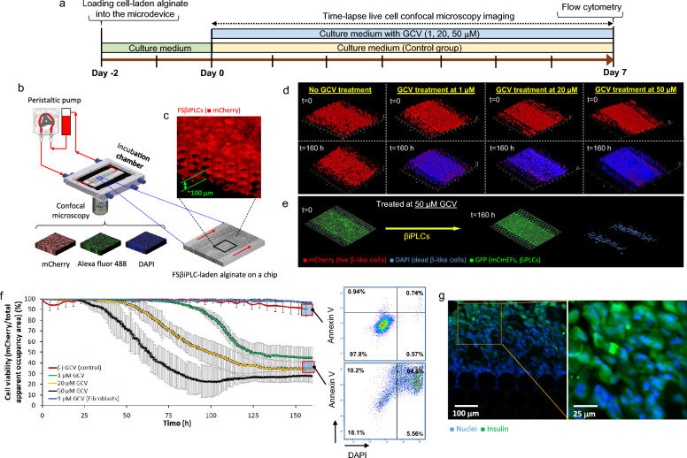
Method: We generated β cell-induced progenitor-like cells (βiPLCs) from primary mouse pancreatic β cells through interrupted reprogramming. Then, we transcriptionally linked our FailSafe (FS) kill-switch, HSV-thymidine kinase (TK), to Cdk1 gene using a CRISPR/Cas9 knock-in strategy, resulting in a FailSafe βiPLC line, designated as FSβiPLCs. Subsequently we evaluated and confirmed the functionality of the drug-inducible kill-switch in FSβiPLCs at different ganciclovir (GCV) concentrations using our PDMS-based transcapillary microfluidic system. Finally, we assessed the functionality of FSβiPLCs by characterizing the dynamics of insulin secretion in response to changes in glucose concentration using our microfluidic perfusion glucose-stimulated insulin secretion (GSIS) assay-on- chip.

Results: The βiPLCs exhibited Ins1, Pdx1 and Nkx6.1 expression, and glucose responsive insulin secretion, the essential properties of pancreatic beta cells. The βiPLCs were amenable to genome editing which allowed for the insertion of the kill-switch into the 3'UTR of Cdk1, confirmed by PCR genotyping. Our transcapillary microfluidic system confirmed the functionality of the drug-inducible kill-switch in FSβiPLCs, showing an effective cell ablation of dividing cells from a heterogeneous cell population at different ganciclovir (GCV) concentrations. The Ki67 expression assessment further confirmed that slow- or non-dividing cells in the FSβiPLC population were resistant to GCV. Our perfusion glucose-stimulated insulin secretion (GSIS) assay-on-chip revealed that the resistant non-dividing FSβiPLCs exhibited higher levels of insulin secretion and glucose responsiveness compared to their proliferating counterparts.

Conclusions: This study establishes a proof-of-concept for the integration of a FailSafe kill-switch system into a glucose-responsive, insulin-secreting cell line to address the safety concerns in stem cell-derived cell replacement treatment for T1D. The microfluidic systems provided valuable insights into the functionality and safety of these engineered cells, demonstrating the potential of the kill-switch to reduce the risk of tumorigenicity in pluripotent cell-derived insulin-secreting cells.

Keywords: Beta cells; Cell reprogramming; Cell therapy; GSIS assay; Microfluidic systems; Organ on chip; Stem cells; Suicide gene; Type 1 diabetes.


Conflict of interest statement

Declarations. Ethics approval and consent to participate: Details of ethics approvals: Title of the approved project: Characterizing the reprogramming process in vivo by using pancreatic endocrine cells to compensate for the beta cell ablated pancreas. Name of the institutional approval committee: The ethics board of The Centre for Phenogenomics (TCP). Approval number: AUP#0182. Date of approval (dd-mm-yy): 08–03-2018. Authors confirm that no human participant was involved in this study; thus, ‘patient consent’ was not applicable for this study. Consent for publication: All authors confirm their consent for publication. Competing interests: The authors declare that they have no competing interests.

Figures

Fig. 1
Fig. 1
Schematics of generation and initial characterization of βiPLCs. a Quadruplet transgenic mice enable On/Off expression of the reprograming Yamanaka factors only in β cells of the pancreas. (See text for more details.) b The experimental design to generate βiPLCs. The relative expression of hallmarks c insulin, d Pdx1 and e Nkx6.1 of βiPLCs compared to the mouse pancreatic islet cells (control), measured by quantitative RT-PCR, f GSIS assay reveals that only line 2 exhibits regulated insulin secretion in response to low and high glucose concentration, g immunostaining of insulin expressed by βiPLCs line 2 (scale bar: 20 μm)
Fig. 2
Fig. 2
a the structure of Failsafe™ knock-in allele of Cdk1 consisting of the suicide gene TK and mCherry reporter compared to the wild type allele of Cdk1. b the representative phase contrast images of FSβiPLCs visualize changes in cell density over 6 days under GCV treatment at 0 (control), 1, 20 and 50 μM GCV (scale bar: 100 μm), c results from the cell viability assay of FSβiPLCs for GCV concentrations of 1, 20 and 50 μM over 10 days compared to the non-treated FSβiPLCs (control), where d GCV treatment (1 μM) leads to the complete ablation of FSmESCs (dividing cells, control), in contrast a certain population of FSβiPLCs survives the GCV treatment which represents the selective ablation of FSβiPLCs, e FSβiPLCs stained with anti-Ki67 antibody (green) and DAPI (blue) to visualize the distribution of Ki67 expressing cells at days 0, 4 and 7 during the GCV treatment of FSβiPLCs at 1, 20 and 50 µM GCV concentrations. f The quantitative evaluation of proliferating and non-proliferating FSβiPLCs represented by flow cytometry analysis of Ki67 protein presentation by FSβiPLCs treated by 1, 20 and 50 µM GCV compared to non-treated FSβiPLCs (control) at days 0, 4 and 7. g immunohistochemistry images show insulin expression by FSβiPLCs corresponding to GCV treatment at 1, 20 and 50 μM in days 0, 4 and 7 compared to non-treated FSβiPLCs (control) (scale bar: 200 μm), h expression of insulin gene by the parental cell line βiPLCs and FSβiPLCs treated at 1, 20 and 50 μM GCV for 7 days as determined by RT-qPCR and normalized to GAPDH relative to insulinoma (MIN6) cell line as the positive control. Astrics denotes statistical significance (**p-value < 0.01, ***p-value < 0.001)
Fig. 3
Fig. 3
a Schematic diagram of the experimental procedure and b set-up for assessing GCV-mediated ablation of FSβiPLCs in a 3D microenvironment on-a-chip, c visualization of the 3D alginate-embedded FSβiPLCs in the microdevice acquired from confocal microscopy imaging, d confocal microscopy images show the dead (DAPI) and living FSβiPLCs (mCherry) and e βiPLCs (GFP) (control) in the 3D microenvironment on-a-chip at the initial (t = 0) and endpoint (t = 160 h) of GCV treatment for different GCV concentrations, f quantitative evaluation of changes and standard deviation (SD) in the apparent cell viability index of FSβiPLCs compared to non-treated FSβiPLCs (control) with time during 7 days of GCV treatment at different GCV concentrations, and represent results from flow cytometry, indicating the live, apoptotic and necrotic cell populations corresponding to the endpoint GCV treatment of the FSβiPLCs on-on-chip at 0 and 20 μM GCV, g in-situ immunohistochemistry of the FSβiPLCs embedded in alginate on-a-chip demonstrates insulin expression by some of FSβiPLCs after 7 days of GCV treatment at 20 μM GCV
Fig. 4
Fig. 4
a Computational simulation of dynamic perfusion GSIS on-a-chip at a flow rate of 10 μL/min shows the velocity field profile, b a mechanistic model for the glucose transportation from the KRB flow in the microchannels into the boundary layer where glucose-uptake and insulin-secretion by the cells take place across the microfluidic device, c a reprehensive computational simulation of insulin concentration distribution across the microfluidic device corresponding to the KRB velocity field at the flow rate of 10 μL/min, d the predict values of temporal changes of insulin concentration at the sampling port, and the anticipated time for the system to reach steady state for sampling, eg a comparison between predicted values from the computational model and experimental data of insulin secretion in terms of normalized insulin secretion per min (%) over time for the GSIS analysis of e βiPLCs, f FSβiPLCs and g GCV treated FSβiPLCs, h measured values of time-dependent insulin content per million of cells for βiPLCs (red) (control), non GCV-treated FSβiPLCs (green) and GCV-treated FSβiPLCs, i the statistical comparison of cumulative insulin content between βiPLCs, FSβiPLCs and the GCV-treated FSβiPLCs at LG (2 mM glucose) and HG (20 mM glucose) where asterisks denote statistically significant differences at any given data point: *p < 0.05, and j the comparison of insulin content between βiPLCs, FSβiPLCs, the GCV-treated FSβiPLCs and MIN6 as a positive control at LG (2 mM) and HG (20 mM)

References

    1. Sugandh F, Chandio M, Raveena F, Kumar L, Karishma F, Khuwaja S, et al. Advances in the management of diabetes mellitus: a focus on personalized medicine. Cureus. 2023;15(8): e43697. - PMC - PubMed
    1. Zakir M, Ahuja N, Surksha MA, Sachdev R, Kalariya Y, Nasir M, et al. Cardiovascular complications of diabetes: from microvascular to macrovascular pathways. Cureus. 2023;15(9): e45835. - PMC - PubMed
    1. Brennan DC, Kopetskie HA, Sayre PH, Alejandro R, Cagliero E, Shapiro AM, et al. Long-Term follow-up of the edmonton protocol of islet transplantation in the United States. Am J Transplant. 2016;16(2):509–17. - PubMed
    1. Shapiro AM, Ricordi C, Hering BJ, Auchincloss H, Lindblad R, Robertson RP, et al. International trial of the Edmonton protocol for islet transplantation. N Engl J Med. 2006;355(13):1318–30. - PubMed
    1. Potter KJ, Westwell-Roper CY, Klimek-Abercrombie AM, Warnock GL, Verchere CB. Death and dysfunction of transplanted β-cells: lessons learned from type 2 diabetes? Diabetes. 2014;63(1):12–9. - PubMed

LinkOut - more resources