. 2024 Dec 19;13(1):64.
doi: 10.1186/s40035-024-00454-5.

CD2AP deficiency aggravates Alzheimer's disease phenotypes and pathology through p38 MAPK activation

Affiliations

CD2AP deficiency aggravates Alzheimer's disease phenotypes and pathology through p38 MAPK activation

Yan-Yan Xue et al. Transl Neurodegener. .

Erratum in

Abstract

Background: Alzheimer's disease (AD) is the most common form of neurodegenerative disorder, which is characterized by a decline in cognitive abilities. Genome-wide association and clinicopathological studies have demonstrated that the CD2-associated protein (CD2AP) gene is one of the most important genetic risk factors for AD. However, the precise mechanisms by which CD2AP is linked to AD pathogenesis remain unclear.

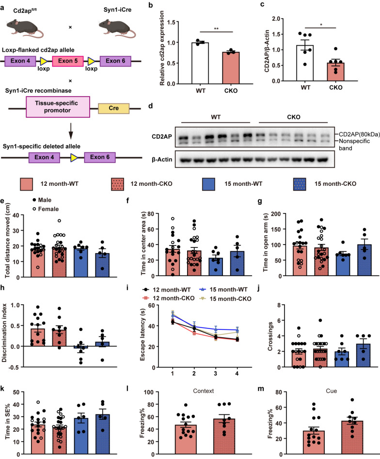
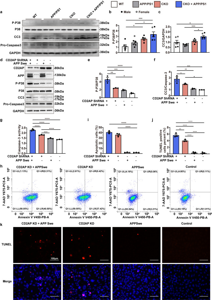
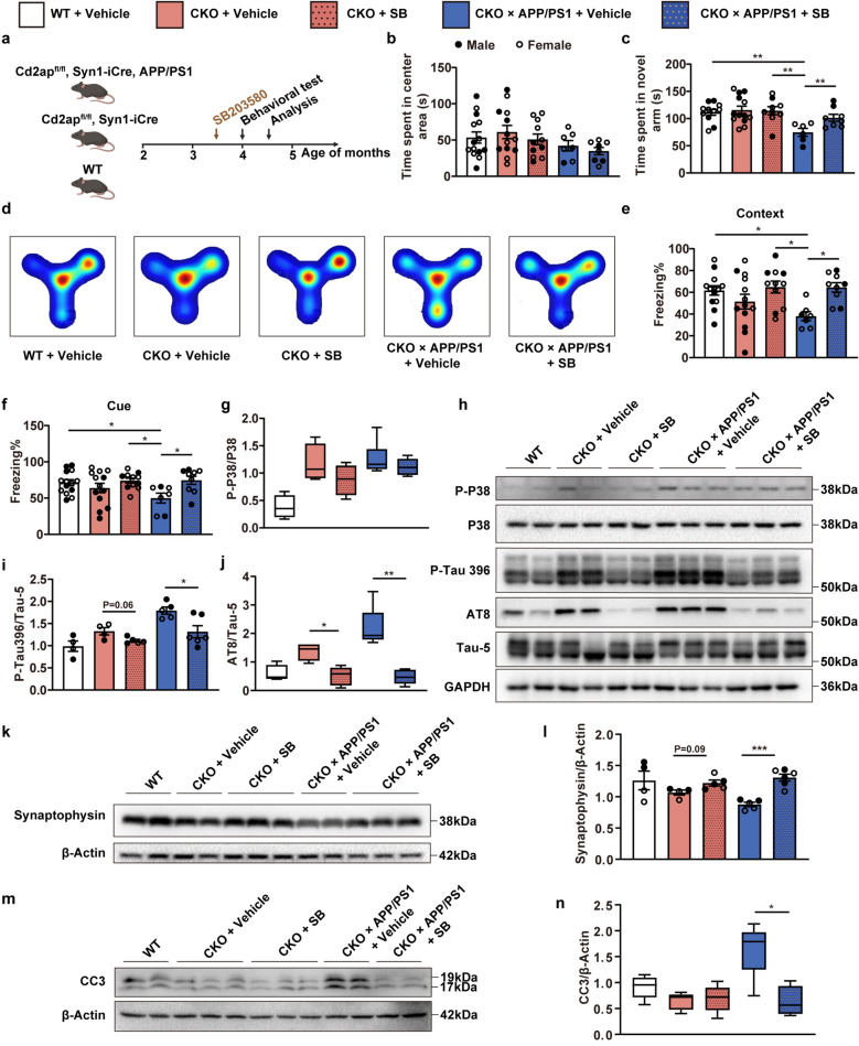
Methods: The spatiotemporal expression pattern of CD2AP was determined. Then, we generated and characterized an APP/PS1 mouse model with neuron-specific Cd2ap deletion, using immunoblotting, immunofluorescence, enzyme-linked immunosorbent assay, electrophysiology and behavioral tests. Additionally, we established a stable CD2AP-knockdown SH-SY5Y cell line to further elucidate the specific molecular mechanisms by which CD2AP contributes to AD pathogenesis. Finally, the APP/PS1 mice with neuron-specific Cd2ap deletion were treated with an inhibitor targeting the pathway identified above to further validate our findings.

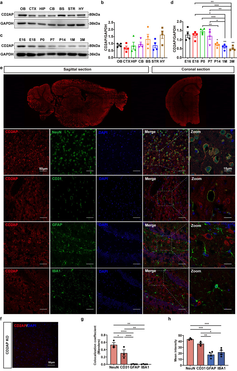
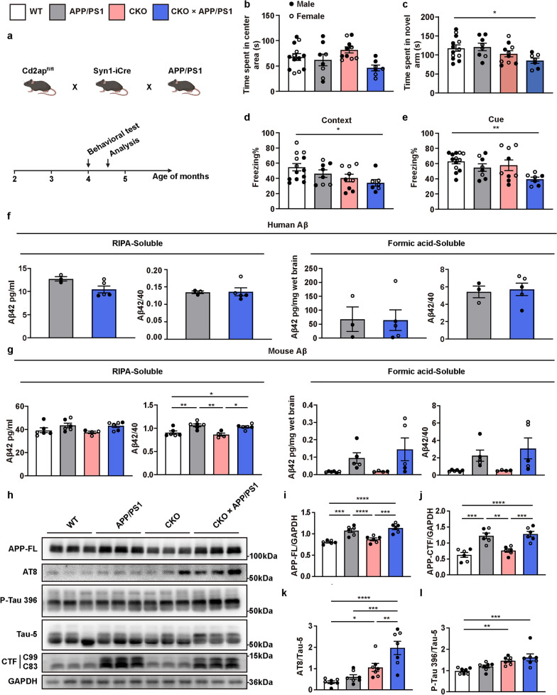
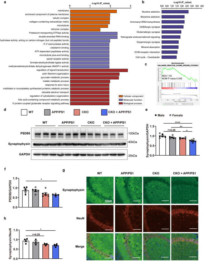
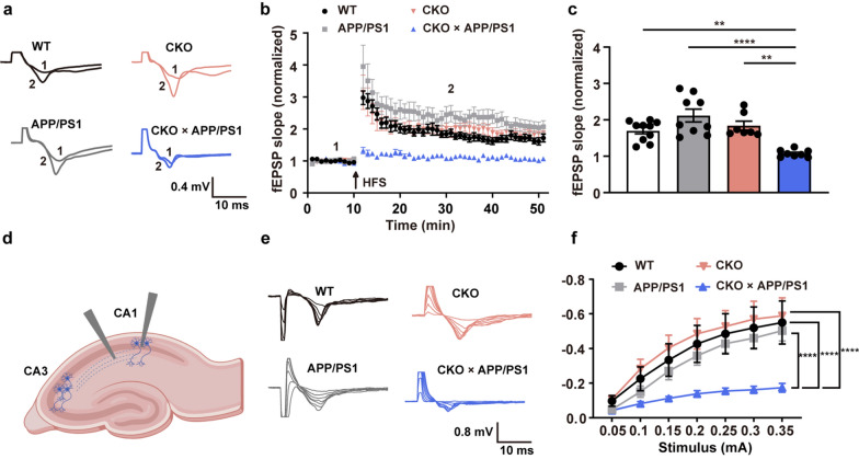
Results: CD2AP is widely expressed in various regions of the mouse brain, with predominant expression in neurons and vascular endothelial cells. In APP/PS1 mice, neuronal knockout of Cd2ap significantly aggravated tau pathology, synaptic impairments and cognitive deficits. Mechanistically, the knockout of Cd2ap activated p38 mitogen-activated protein kinase (MAPK) signaling, which contributed to increased tau phosphorylation, synaptic injury, neuronal apoptosis and cognitive impairment. Furthermore, the phenotypes of neuronal Cd2ap knockout were ameliorated by a p38 MAPK inhibitor.

Conclusion: Our study presents the first in vivo evidence that CD2AP deficiency exacerbates the phenotypes and pathology of AD through the p38 MAPK pathway, identifying CD2AP/p38 MAPK as promising therapeutic targets for AD.

Keywords: Alzheimer’s disease; CD2AP; P38 MAPK; Synaptic injury; Tau.


Conflict of interest statement

Declarations. Ethics approval and consent to participate: Animal experiments were approved by the Institute of Animal Care Committee of the Second Affiliated Hospital, Zhejiang University School of Medicine (No. 202036). Consent for publication: Not applicable. Competing interest: The authors declare no competing interests. Author disclosures are available in the supporting information.

Figures

Fig. 1
Fig. 1
Expression of CD2AP in the brains of mice. a, b Immunoblots showed protein expression of CD2AP in the different mouse brain regions of 3-month-old mice. OB, Olfactory bulb; CTX, Cerebral cortex; HIP, hippocampus; CB, cerebellum; BS, brainstem; STR, striatum; HY, hypothalamus. c, d Immunoblots showed protein expression of CD2AP in whole-brain lysates from mice at the indicated stages of development. e Representative confocal images for CD2AP, NEUN, CD31, GFAP, IBA1 and DAPI in mouse cortex and hippocampus from 3-month-old mice. f Immunofluorescence image of CD2AP in the 1-month-old KO mouse brain as a negative control. g Quantitative analysis of fluorescence colocalization using Mander’s overlap coefficient methods. h The mean fluorescence intensity of CD2AP within the boundaries of the different cellular markers. All data are presented as mean ± SEM. One-way ANOVA with Turkey’s multiple comparison tests for multiple comparisons (b, d, g, h). *P < 0.05, **P < 0.01
Fig. 2
Fig. 2
Neuronal Cd2ap deletion mice were not cognitively impaired. a Scheme of the generation of a conditional Cd2ap knockout mouse line. b RT-PCR analysis of CD2AP mRNA levels in whole-brain lysates from 12-month-old WT and CKO mice. c, d Immunoblots showing decreased protein expression of CD2AP in the brains of 12-month-old CKO mice compared to age-matched WT mice. e, f No significant difference in total distance moved or time spent in the center area was observed in the open field test. n = 18 (12 month-WT, female n = 5, male n = 13), n = 22 (12 month-CKO, female n = 13, male n = 9), n = 7 (15 month-WT, all male), n = 5 (15 month-CKO, all males). g No significant difference in time spent in the open arm was observed in the elevated plus maze. n = 18 (12 month-WT, female n = 5, male n = 13), n = 22 (12 month-CKO, female n = 13, male n = 9), n = 7 (15 month-WT, all males), n = 5 (15 month-CKO, all males). h There was no difference in preference for the novel object between WT and CKO both at 12 months and 15 months. n = 13 (12 month-WT, all male), n = 9 (12 month-CKO, all male), n = 7 (15 month-WT, all male), n = 5 (15 month-CKO, all males). i-k In the Morris Water maze, CKO and WT mice showed no difference in escape latency, the number of crossings of the platform or time spent in the target area. n = 18 (12 month-WT, female n = 5, male n = 13), n = 22 (12 month-CKO, female n = 13, male n = 9), n = 7 (15 month-WT), n = 5 (15 month-CKO). l, m In the fear conditioning test, 12-month-old CKO mice showed a similar freezing time to WT mice. n = 14 (WT, all male), n = 9 (CKO, all male). All data are presented as mean ± SEM. Unpaired t-test with two-tailed analysis was conducted separately for 12- and 15-month-old mice. *P < 0.05, **P < 0.01
Fig. 3
Fig. 3
Neuronal Cd2ap deletion aggravated cognitive function and pathological features in APP/PS1 mice. a Scheme of the experimental mouse timeline. Briefly, a series of behavioral tests were performed in 4-month-old mice, and subsequent pathological analyses were conducted in 4.5-month-old mice. b No significant difference in time spent in the center area was observed in the open field test. n = 13 (WT, female n = 5, male n = 8), n = 8 (APP/PS1, female n = 3, male n = 5), n = 10 (CKO, female n = 5, male n = 5), n = 7 (CKO × APP/PS1, female n = 4, male n = 3). c CKO × APP/PS1 mice spent less time in the novel arm in the Y-maze novel arm preference test compared to WT mice. n = 13 (WT, female n = 5, male n = 8), n = 8 (APP/PS1, female n = 3, male n = 5), n = 10 (CKO, female n = 5, male n = 5), n = 7 (CKO × APP/PS1, female n = 4, male n = 3). d, e CKO × APP/PS1 mice showed significantly decreased contextual and cue-related freezing compared to WT mice. n = 13 (WT, female n = 5, male n = 8), n = 8 (APP/PS1, female n = 3, male n = 5), n = 9 (CKO, female n = 5, male n = 4), n = 7 (CKO × APP/PS1, female n = 4, male n = 3). f ELISA analysis of Aβ showed that neuronal Cd2ap deletion had no obvious influence on human Aβ level. n = 3 (APP/PS1, female n = 1, male n = 2), n = 5 (CKO × APP/PS1, female n = 3, male n = 2). g ELISA analysis of Aβ showed that neuronal Cd2ap deletion had no obvious influence on murine Aβ level. n = 6 (WT, female n = 2, male n = 4), n = 6 (APP/PS1, female n = 3, male n = 3), n = 4 (CKO, female n = 3, male n = 1), n = 6 (CKO × APP/PS1, female n = 4, male n = 2). h-j In 4.5-month-old mice, Immunoblots revealed that neuronal Cd2ap deletion had no obvious influence on the full-length APP (APP-FL) and APP-CTF proteins. n = 6 (WT, female n = 3, male n = 3), n = 6 (APP/PS1, female n = 3, male n = 3), n = 6 (CKO, female n = 3, male n = 3), n = 6 (CKO × APP/PS1, female n = 3, male n = 3). k, l In 4.5-month-old mice, immunoblots revealed that neuronal Cd2ap deletion led to significantly increased ptau202/205 (AT8) and p-tau396 level, especially in the CKO × APP/PS1 mice. n = 7 (WT, female n = 3, male n = 4), n = 7 (APP/PS1, female n = 3, male n = 4), n = 7 (CKO, female n = 4, male n = 3), n = 7 (CKO × APP/PS1, female n = 4, male n = 3). All data are presented as mean ± SEM. Unpaired t-test with two-tailed analysis (f), one-way ANOVA with Turkey’s multiple comparison tests for multiple comparisons (b-e, g, i, j, k), Kruskal–Wallis tests with Dunn’s multiple comparison tests (l). *P < 0.05, **P < 0.01, ***P < 0.001, ****P < 0.0001
Fig. 4
Fig. 4
Proteomic analyses in Cd2ap knockout mice and neuronal Cd2ap deletion led to synaptic loss. a GO annotation analysis based on differentially expressed proteins was presented as the top six in cellular component (CC), and the top ten in biological process (BP) and molecular function (MF). b The top ten enriched pathways from KEGG pathway analysis of differentially expressed proteins. c Gene set enrichment analysis (GSEA) revealed that the reactive oxygen species pathway was upregulated in the Cd2ap knockout group. d, e Immunoblots showed a decreased synaptophysin level in the 4.5-month-old CKO and CKO × APP/PS1 mice. n = 6 (WT, female n = 3, male n = 3), n = 6 (APP/PS1, female n = 3, male n = 3), n = 5 (CKO, female n = 3, male n = 2), n = 7 (CKO × APP/PS1, female n = 4, male n = 3). f Immunoblots showed a decreased PSD95 level in the 4.5-month-old CKO × APP/PS1 mice. n = 6 (WT, female n = 3, male n = 3), n = 6 (APP/PS1, female n = 3, male n = 3), n = 6 (CKO, female n = 3, male n = 3), n = 5 (CKO × APP/PS1, female n = 2, male n = 3). g, h Immunofluorescence analysis showed significantly decreased synaptophysin in the hippocampus in 4.5-month-old CKO and CKO × APP/PS1 mice. n = 4 (WT, all male), n = 3 (APP/PS1, all male), n = 3 (CKO, all male), n = 4 (CKO × APP/PS1, all male). All data are presented as mean ± SEM. One-way ANOVA with Turkey’s multiple comparison tests for multiple comparisons (e, h), Kruskal–Wallis tests with Dunn’s multiple comparison tests (f). *P < 0.05, **P < 0.01, ***P < 0.001, ****P < 0.0001
Fig. 5
Fig. 5
Neuronal Cd2ap deletion led to synaptic deficits in 4.5-month-old APP/PS1 mice. a Representative traces of fEPSP during LTP recording. b Time-course of fEPSP slopes during LTP recording. c The normalized average fEPSP slopes during the last 10 min. n = 10 from 3 mice in WT group, n = 9 from 3 mice in APP/PS1 group, n = 7 from 3 mice in CKO group, n = 8 from 3 mice in CKO × APP/PS1 group, one-way ANOVA with Turkey’s multiple comparison tests for multiple comparisons. d Schematic diagram showing the localization of stimulating and recording electrodes in the hippocampal slice. e Representative I/O curves for fEPSPs recorded in hippocampal CA1 subregion from 4 groups. f Quantitative analyses of I/O curves. n = 10 from 3 male mice in WT group, n = 9 from 3 male mice in APP/PS1 group, n = 9 from 3 male mice in CKO group, n = 9 from 3 male mice in CKO × APP/PS1 group, two-way ANOVA. HFS: High-frequency stimulation. All data are presented as mean ± SEM. *P < 0.05, **P < 0.01, ***P < 0.001, ****P < 0.0001
Fig. 6
Fig. 6
Neuronal Cd2ap knockout activated p38 MAPK signaling and apoptosis. ac Immunoblots showed significantly increased phosphorylated p38 MAPK in 4.5-month-old CKO and CKO × APP/PS1 mice and significantly increased CC3 level in 4.5-month-old CKO × APP/PS1 mice. n = 6 (WT, female n = 3, male n = 3), n = 6 (APP/PS1, female n = 3, male n = 3), n = 6 (CKO, female n = 3, male n = 3), n = 6 (CKO × APP/PS1, female n = 3, male n = 3). df Immunoblots showed significantly increased p-p38 MAPK and CC3 levels in CD2AP stable knockdown cells and especially in KD cells with APPSwe co-expression. g Knockdown of CD2AP with APPSwe expression significantly increased caspase-3 activity in SH-SY5Y cells. h, i Analysis of flow cytometry showed that the number of apoptotic cells was significantly increased in CD2AP knockdown cells and especially in CD2AP knockdown cells with APPSwe co-expression. The vertical axis represented the cells labeled with 7-AAD and the horizontal axis indicated those stained with Annexin V (eFluor™ 450). The percentage of cells labeled with Annexin V but without 7-AAD was calculated and compared. j, k The apoptotic cells were detected with the TUNEL assay with nuclei stained with DAPI. The percentage of cells was quantified in different groups. All data are presented as mean ± SEM, n = 3 per group (dk). One-way ANOVA with Turkey’s multiple comparison test for multiple comparisons. *P < 0.05, **P < 0.01, ***P < 0.001, ****P < 0.0001
Fig. 7
Fig. 7
Inhibition of p38 MAPK rescued learning deficit, synaptic loss, and apoptosis in CKO × APP/PS1 mice. a Scheme of drug treatment and test schedules. b No significant difference in time spent in the center area was observed among 4-month-old mice in the open field test. n = 14 (WT, female n = 8, male n = 6), n = 13 (CKO + Vehicle, female n = 6, male n = 7), n = 11 (CKO + SB, female n = 7, male n = 4), n = 7 (CKO × APP/PS1 + Vehicle, female n = 5, male n = 2), n = 8 (CKO × APP/PS1 + SB, female n = 5, male n = 3). c, d In the Y-maze novel arm preference test, 4-month-old CKO × APP/PS1 mice given SB203580 treatment spent more time in the novel arm than those given the vehicle. n = 11 (WT, female n = 7, male n = 4), n = 13 (CKO + Vehicle, female n = 6, male n = 7), n = 9 (CKO + SB, female n = 6, male n = 3), n = 6 (CKO × APP/PS1 + Vehicle, female n = 4, male n = 2), n = 8 (CKO × APP/PS1 + SB, female n = 6, male n = 2). e, f Four-month-old CKO × APP/PS1 mice given SB203580 treatment showed significantly increased contextual and cue-related freezing time than those given the vehicle. n = 14 (WT, female n = 8, male n = 6), n = 13 (CKO + Vehicle, female n = 6, male n = 7), n = 11 (CKO + SB, female n = 7, male n = 4), n = 7 (CKO × APP/PS1 + Vehicle, female n = 5, male n = 2), n = 9 (CKO × APP/PS1 + SB, female n = 6, male n = 3). g SB203580 treatment did not obviously suppress the phosphorylated p38 MAPK in 4.5-month-old CKO and CKO × APP/PS1 mice. n = 4 (WT, female n = 2, male n = 2), n = 4 (CKO + Vehicle, female n = 2, male n = 2), n = 4 (CKO + SB, female n = 2, male n = 2), n = 6 (CKO × APP/PS1 + Vehicle, female n = 4, male n = 2), n = 6 (CKO × APP/PS1 + SB, female n = 3, male n = 3) hj SB203580 treatment significantly decreased the hyperphosphorylation of tau in 4.5-month-old CKO and CKO × APP/PS1 mice, n = 4 (WT, female n = 2, male n = 2), n = 4 (CKO + Vehicle, female n = 2, male n = 2), n = 5 (CKO + SB, female n = 2, male n = 3), n = 5 (CKO × APP/PS1 + Vehicle, female n = 3, male n = 2), n = 6 (CKO × APP/PS1 + SB, female n = 3, male n = 3). k, l SB203580 treatment increased synaptophysin protein level in 4.5-month-old CKO and CKO × APP/PS1 mice. n = 4 (WT, female n = 2, male n = 2), n = 4 (CKO + Vehicle, female n = 2, male n = 2), n = 5 (CKO + SB, female n = 2, male n = 3), n = 5 (CKO × APP/PS1 + Vehicle, female n = 3, male n = 2), n = 6 (CKO × APP/PS1 + SB, female n = 3, male n = 3). m, n SB203580 treatment significantly decreased CC3 level in 4.5-month-old CKO × APP/PS1 mice. n = 5 (WT, female n = 3, male n = 2), n = 5 (CKO + Vehicle, female n = 3, male n = 2), n = 5 (CKO + SB, female n = 2, male n = 3), n = 5 (CKO × APP/PS1 + Vehicle, female n = 3, male n = 2), n = 4 (CKO × APP/PS1 + SB, female n = 2, male n = 2). Data are presented as mean ± SEM (b, c, e f, i, l) and box plots with median (g, j, n). One-way ANOVA with Turkey’s multiple comparison tests for multiple comparisons (b-f), One-way ANOVA or Kruskal–Wallis tests for detecting overall differences among multiple groups (g-n), followed by specific comparisons, including CKO + Vehicle vs. CKO + SB, and CKO × APP/PS1 + Vehicle vs. CKO × APP/PS1 + SB, using unpaired t-test with two-tailed analysis (i, l) and Mann–Whitney test (g, j, n), with Bonferroni correction applied. *P < 0.05, **P < 0.01, ***P < 0.001

References

    1. Tao QQ, Cai X, Xue YY, Ge W, Yue L, Li XY, et al. Alzheimer’s disease early diagnostic and staging biomarkers revealed by large-scale cerebrospinal fluid and serum proteomic profiling. Innovation. 2024;5:100544. - PMC - PubMed
    1. Kunkle BW, Schmidt M, Klein H-U, Naj AC, Hamilton-Nelson KL, Larson EB, et al. Novel Alzheimer disease risk loci and pathways in african american individuals using the african genome resources panel: a meta-analysis. JAMA Neurol. 2021;78:102–13. - PMC - PubMed
    1. Hollingworth P, Harold D, Sims R, Gerrish A, Lambert J-C, Carrasquillo MM, et al. Common variants at ABCA7, MS4A6A/MS4A4E, EPHA1, CD33 and CD2AP are associated with Alzheimer’s disease. Nat Genet. 2011;43:429–35. - PMC - PubMed
    1. Naj AC, Jun G, Beecham GW, Wang L-S, Vardarajan BN, Buros J, et al. Common variants at MS4A4/MS4A6E, CD2AP, CD33 and EPHA1 are associated with late-onset Alzheimer’s disease. Nat Genet. 2011;43:436–41. - PMC - PubMed
    1. Chen H, Wu G, Jiang Y, Feng R, Liao M, Zhang L, et al. Analyzing 54,936 samples supports the association between CD2AP rs9349407 Polymorphism and Alzheimer’s Disease susceptibility. Mol Neurobiol. 2015;52:1–7. - PubMed

Publication types

Substances

LinkOut - more resources