. 2025 Apr;12(2):1304-1315.
doi: 10.1002/ehf2.15145. Epub 2024 Dec 18.

Sacubitril/valsartan preserves regional cardiac function following myocardial infarction in rats

Affiliations

Sacubitril/valsartan preserves regional cardiac function following myocardial infarction in rats

Einar Sjaastad Nordén et al. ESC Heart Fail. 2025 Apr.

Abstract

Aims: Sacubitril/valsartan (Sac/Val) is used for treatment of heart failure. The effect of Sac/Val on regional dysfunction following myocardial infarction (MI) remains uncertain. This study aimed at understanding the effects of Sac/Val on regional function after MI.

Methods and results: MI or sham surgery was performed in Sprague-Dawley rats. Animals were randomized to treatment with Sac/Val, valsartan (Val) or vehicle (Veh). Magnetic resonance imaging was used to acquire left ventricular volumes and strain. Left ventricular tissue was obtained for wesern blotting, PCR and Masson's trichrome staining. Isolated cardiac fibroblasts were cultured with Veh, atrial natriuretic peptide (ANP), adrenomedullin (ADM) and sacubitrilat, and collagen expression assessed with droplet digital PCR.

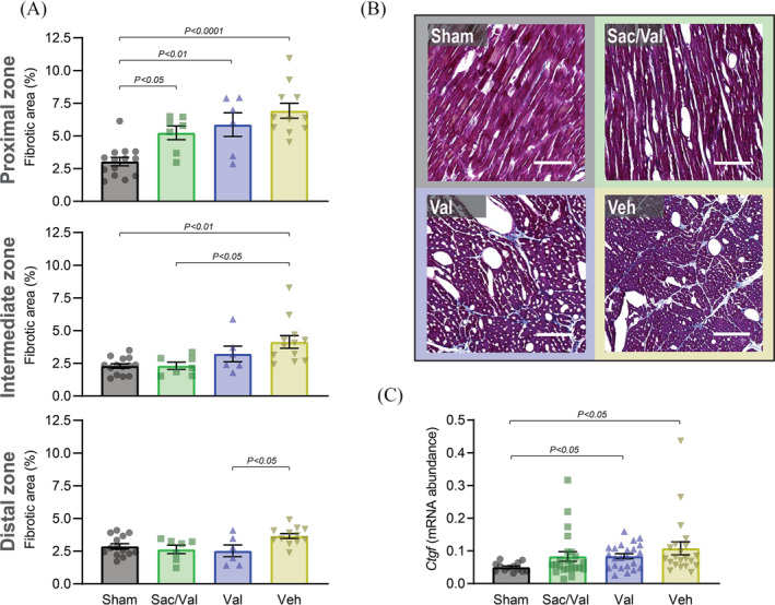
Results: Sac/Val reduced ventricular end-diastolic volume by 18% compared with Veh, and preserved circumferential systolic strain in the zone proximal to infarction compared with sham after 42 days of treatment (peak strain ± SEM: sham: -0.19 ± 0.01%; Sac/Val: -0.14 ± 0.02%; Val: -0.10 ± 0.02%; Veh: -0.10 ± 0.02%). Masson's trichrome staining demonstrated lower fibrotic deposition in the intermediate zone with Sac/Val treatment than Veh (sham: 2.29 ± 0.17%; Sac/Val: 2.31 ± 0.27%; Val: 3.22 ± 0.60%; Veh: 4.14 ± 0.48%). The amounts of the pro-apoptotic caspase 3 cleavage fragments p19/17 were 89% higher in Val than sham, with Sac/Val showing no significant increase compared with sham. Collagen expression in human fibroblast culture was lower in cells co-treated with sacubitrilat and ANP, an effect not observed with sacubitrilat/ADM co-treatment.

Conclusions: Sac/Val preserves in vivo myocardial function in the region most proximal to MI in rats and reduces left ventricular dilatation. These effects may be related to a reduction in both fibrosis and pro-apoptotic signalling.

Keywords: ARNi; Heart failure; Myocardial infarction; Regional function; Sacubitril/valsartan.


Conflict of interest statement

Sacubitril/valsartan and valsartan utilized for in vivo studies were received free of charge from Novartis AG. Dr. Hussain was an employee of Novartis AG at time of study conduct. The other authors declare that they do not have any conflict of interest.

Figures

Figure 1
Figure 1
Sacubitril/valsartan treatment decreases left ventricular dilatation. (A) Graphical representation of study design. After surgery and drug treatment, animals underwent imaging and tissue harvesting at days 10, 21 and 42. Tissues from a subset of animals were acquired at day 3. Sac/Val: sacubitril/valsartan. Val: valsartan. Veh: vehicle. (B) Left panel: MRI‐based measurement of ventricular ejection fraction (LVEF). (Day 10: sham n = 12; Sac/Val n = 9; Val n = 4; Veh n = 13. Day 21: sham n = 10, Sac/Val n = 4; Val n = 4; Veh n = 6. Day 42: sham n = 26; Sac/Val n = 12; Val n = 6; Veh n = 20). Right panel: MRI‐based measurement of left ventricular end‐diastolic volume (LVEDV) (n same as LVEF). Welch ANOVA, multiple comparison between all groups, significant differences are shown. LAD, left anterior descending artery. Parts of the figure were drawn by using pictures from Servier Medical Art. Servier Medical Art by Servier is licensed under a Creative Commons Attribution 3.0 Unported Licence (https://creativecommons.org/licenses/by/3.0/).
Figure 2
Figure 2
Sacubitril/valsartan treatment preserves systolic function in the proximal zone. (A) MRI based measurement of circumferential systolic strain in proximal, intermediate, and distal zones at day 42. Sac/Val: Sacubitril/Valsartan. Val: Valsartan. Veh: Vehicle. (B) Representative strain curves from MRI tissue phase mapping imaging derived circumferential strain. Data for sham represent global circumferential strain. (C) Representative mid‐left‐ventricular CINE MRI images, with 3 mm white scale bar, from 1: sham, 2: Sac/Val, 3: Val, 4: Veh. Left images are end‐diastole and right images are end‐systole. Welch ANOVA, multiple comparison between all groups, significant differences are shown.
Figure 3
Figure 3
Sacubitril/valsartan treatment reduces fibrosis in the intermediate zone. (A) Masson's trichrome staining of mid‐ventricular sections for quantification of fibrotic area with quantification of fibrotic deposition in the proximal, intermediate, and distal zone. Sac/Val: Sacubitril/Valsartan. Val: Valsartan. Veh: Vehicle. (B) Representative images from Masson's trichrome stained sections from the intermediate zone. Blue represents fibrosis and red cardiomyocytes. Scale bar is 50 μm. Adjustment of contrast to match reference image have been applied in all images. (C) ddPCR measurement of Ctgf in the intermediate zone on day 42. One‐way ANOVA, multiple comparison between all groups, significant differences are shown.
Figure 4
Figure 4
Sacubitril/valsartan treatment promotes an anti‐apoptotic profile compared with valsartan alone. (A) ddPCR (top) and western blotting (bottom) for BAX in tissue from the proximal zone at day 10 (ddPCR: One‐way ANOVA, western blotting: Kruskal–Wallis). Sac/Val: sacubitril/valsartan. val: valsartan. Veh: vehicle. (B) Western blotting of tissue from proximal zone at day 10 for caspase 3 (top) and caspase 3 cleavage fragment p19/17 (bottom) (Kruskal–Wallis). (C) ddPCR (top) and western blotting (bottom) from the proximal zone at day 10 for BCL‐2 (one‐way ANOVA). Multiple comparison between all groups, significant differences are shown.
Figure 5
Figure 5
In vitro expression of collagen is reduced by treatment with sacubitril/valsartan. qRT‐PCR of Col1a2 from cell culture of human cardiac fibroblasts with 16 h co‐culture with drug or hormone treatment. Experiments were performed in triplicate, with two biological replicates per run. (A) Vehicle, atrial natriuretic peptide (ANP) or adrenomedullin (ADM) added to culture. (B) Vehicle, Sacubitrilat (Sac), Sac combined with ANP, or Sac combined with ADM added to culture. Nested one‐way ANOVA, multiple comparison towards Vehicle, significant differences are shown.
Figure 6
Figure 6
Treatment with Sacubitril/Valsartan reduces calcium transient amplitude. (A) Western blotting of left ventricular tissue from proximal zone. Top panel: SERCA2. Bottom panel: phospholamban (PLB). Sac/Val: sacubitril/valsartan. Val: valsartan. Veh: vehicle. One‐way ANOVA, multiple comparison between all groups, significant differences are shown. (B) Confocal microscopy line‐scans of isolated cardiomyocytes stimulated with various drugs. Values normalized to Vehicle. Repeats given are biological repeats Left panels: Calcium transient amplitude. Top: Stimulation of cells with either vehicle (n = 4), sacubitrilat (Sac) (n = 3) or valsartan (Val) (n = 4). Bottom: Stimulation of cells with either vehicle (n = 5), Sac combined with ANP (n = 3), or Sac and Val in combination with ANP (n = 3). Right panels: Spontaneous Ca2+ spark frequency. Top: Stimulation of cells with either vehicle (n = 4), sacubitrilat (Sac) (n = 3) or valsartan (Val) (n = 4). Bottom: Stimulation of cells with either vehicle (n = 5), Sac combined with ANP (n = 3), or Sac and Val in combination with ANP (n = 3). Nested one‐way ANOVA, multiple comparison towards vehicle, significant differences are shown. (C) Confocal microscopy line scans demonstrating calcium transient characteristics in isolated cardiomyocytes stimulated with 1 Hz pulses, representative transients from each treatment condition shown in panel (B). Scale bar represents 1 s.

References

    1. McMurray JJ, Packer M, Desai AS, Gong J, Lefkowitz MP, Rizkala AR, et al. Angiotensin‐neprilysin inhibition versus enalapril in heart failure. N Engl J Med 2014;371:993‐1004. doi:10.1056/NEJMoa1409077 - DOI - PubMed
    1. Ziaeian B, Fonarow GC. Epidemiology and aetiology of heart failure. Nat Rev Cardiol 2016;13:368‐378. doi:10.1038/nrcardio.2016.25 - DOI - PMC - PubMed
    1. Perera D, Clayton T, O'Kane PD, Greenwood JP, Weerackody R, Ryan M, et al. Percutaneous revascularization for ischemic left ventricular dysfunction. N Engl J Med 2022;387:1351‐1360. doi:10.1056/NEJMoa2206606 - DOI - PubMed
    1. Pfeffer MA, Claggett B, Lewis EF, Granger CB, Køber L, Maggioni AP, et al. Angiotensin receptor–neprilysin inhibition in acute myocardial infarction. N Engl J Med 2021;385:1845‐1855. doi:10.1056/NEJMoa2104508 - DOI - PubMed
    1. Rezq A, Saad M, El Nozahi M. Comparison of the efficacy and safety of sacubitril/valsartan versus ramipril in patients with ST‐segment elevation myocardial infarction. Am J Cardiol 2021;143:7‐13. doi:10.1016/j.amjcard.2020.12.037 - DOI - PubMed

MeSH terms

LinkOut - more resources