. 2024 Dec;4(12):e70079.
doi: 10.1002/cpz1.70079.

Development of a Microphysiological Cartilage-on-Chip Platform for Dynamic Biomechanical Stimulation of Three-Dimensional Encapsulated Chondrocytes in Agarose Hydrogels

Affiliations

Development of a Microphysiological Cartilage-on-Chip Platform for Dynamic Biomechanical Stimulation of Three-Dimensional Encapsulated Chondrocytes in Agarose Hydrogels

Valtteri Peitso et al. Curr Protoc. 2024 Dec.

Abstract

Osteoarthritis (OA) is one of the most prevalent joint diseases globally, characterized by the progressive breakdown of articular cartilage, resulting in chronic pain, stiffness, and loss of joint function. Despite its significant socioeconomic impact, therapeutic options remain limited, largely due to an incomplete understanding of the molecular mechanisms driving cartilage degradation and OA pathogenesis. Recent advances in in vitro modeling have revolutionized joint tissue research, transitioning from simplistic two-dimensional cell cultures to sophisticated three-dimensional (3D) constructs that more accurately mimic the physiological microenvironment of native cartilage. Over the last decade, organ-on-chip technologies have emerged as transformative tools in tissue engineering, offering microphysiological platforms with precise control over biomechanical and biochemical stimuli. These platforms are providing novel insights into tissue responses and disease progression and are increasingly integrated into the early stages of drug screening and development. In this article, we present a detailed experimental protocol for constructing a cartilage-on-chip system capable of delivering controlled dynamic biomechanical stimulation to 3D-encapsulated chondrocytes in an agarose hydrogel matrix. Our protocol, optimized for both bovine and human chondrocytes, begins with Basic Protocol 1, detailing the preparation and injection of cell-laden hydrogels into the microdevice. Basic Protocol 2 describes the application of dynamic mechanical loading using a calibrated pressurized pump. Finally, Basic Protocols 3 and 4 focus on the retrieval of the hydrogel and RNA extraction for downstream molecular analyses. This platform represents a critical advancement for in vitro studies of cartilage biology, enabling more precise modeling of OA pathophysiology and evaluation of experimental therapeutics. © 2024 The Author(s). Current Protocols published by Wiley Periodicals LLC. Basic Protocol 1: Cartilage-on-chip injection Basic Protocol 2: Cartilage-on-chip actuation Basic Protocol 3: Cartilage-on-chip agarose hydrogel removal Basic Protocol 4: Preparation of cartilage-on-chip for RNA extraction.

Keywords: cartilage; chondrocyte; microphysiological; organ‐on‐a‐chip; osteoarthritis.


Conflict of interest statement

Carlo Alberto Paggi and Elsa Lauwers work for chrn on‐chip biotechnology B.V. and have helped in the protocol drafting.

Figures
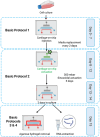
Figure 1
Figure 1
A representative overview of the workflow described in this protocol. Days 0‐7: Basic Protocol 1 involves cartilage‐on‐chip injection and maintenance with the medium being changed every 2 days. Days 8‐12: Basic Protocol 2 focuses on cartilage‐on‐chip actuation, where 300 mbar sinusoidal pressure is applied for 1 hr every day for the duration of 5 days. Days 13‐14: System in culture without actuation. Day 15: Depending on downstream analysis either Basic Protocol 3 or 4 is performed. The figure was created using BioRender.
Figure 2
Figure 2
Illustrative figure of the cartilage‐on‐chip device containing the actuation chamber, cell chamber, and perfusion channel. The figure was created using BioRender.
Figure 3
Figure 3
Illustrative figure of the injection of the cell‐agarose‐suspension into the cell chamber of the cartilage‐on‐chip device. The figure was created using BioRender.
Figure 4
Figure 4
Illustrative figure of the second injection of the cell‐agarose‐suspension into the cell chamber of the cartilage‐on‐chip device. The figure was created using BioRender.
Figure 5
Figure 5
Illustrative figure of the injection of the complete medium into the perfusion channel of the cartilage‐on‐chip device. The figure was created using BioRender.
Figure 6
Figure 6
Chip illustrations under the microscope. Left: Schematic view of the chip and the area seen under the microscope. Middle: The whole view of the chip under the microscope shows the mechanical actuation unit, cell‐agarose‐hydrogel, and perfusion channel. Right: Close‐up view of cells embedded in agarose hydrogel under different conditions: Static, 300 mbar, and 700 mbar pressure (cells shown by red arrows; scale bar = 15 µm).
Figure 7
Figure 7
Calibration of the pressure controller instrument. Red arrows indicate the buttons to be pressed.
Figure 8
Figure 8
Selection of the correct waveform in the pressure controller instrument. Red arrows indicate the buttons to be pressed.
Figure 9
Figure 9
Setting the pressure and the cyclic period in the pressure controller instrument. Red arrows indicate the buttons to be pressed.
Figure 10
Figure 10
Replacing Cartila‐plate lid with Cartila‐plate lid with holes on Cartila‐plate.
Figure 11
Figure 11
Insertion of the tubing into the hole of the actuation chamber of the cartilage‐on‐chip device. (A) An illustrative figure of the actuation chamber. (BC) Insertion of the tubing into the actuation channel.
Figure 12
Figure 12
Representation of the (A) actuation chamber and (B) Cartila‐plate with cartilage‐on‐chip devices, tubing, and Cartila‐plate lid with holes.
Figure 13
Figure 13
The actuation system containing a pressure controller instrument, 7‐port connector, Cartila‐plate with cartilage‐on‐chip devices, Cartila‐plate lid with holes, tubing, and connectors.
Figure 14
Figure 14
Removal of the transparent section of the cartilage‐on‐chip device from the single unit holder.
Figure 15
Figure 15
Cutting the transparent section of the cartilage‐on‐chip device with a razor blade. (A) An illustration of the cutting sites. (BD) Illustrations of the cutting of the transparent sections. (E) A cartilage‐on‐chip device cut from the three sides.
Figure 16
Figure 16
Cutting the transparent section of the cartilage‐on‐chip device's actuation channel with a scalpel. (A) Insertion of the blade into the actuation unit hole. (B) Illustration of the cut along the actuation unit.
Figure 17
Figure 17
Removal of the agarose hydrogel from the transparent part of the cartilage‐on‐chip device.
Figure 18
Figure 18
Removal of the liquid with a tissue wipe from the agarose hydrogel.
Figure 19
Figure 19
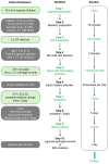
A flowchart of critical parameters, workflow, and duration. The figure was created using BioRender.

References

Literature Cited

    1. Banh, L. , Cheung, K. K. , Chan, M. W. Y. , & Young, E. W. K. (2022). Viswanathan S. Advances in organ‐on‐a‐chip systems for modelling joint tissue and osteoarthritic diseases. Osteoarthritis Cartilage, 30(8), 1050–1061. 10.1016/j.joca.2022.03.012 - DOI - PubMed
    1. Buckwalter, J. A. , Mankin, H. J. , & Grodzinsky, A. J. (2005). Articular cartilage and osteoarthritis. Instructional Course Lectures, 54, 465–80. - PubMed
    1. Buckwalter, J. A. , Martin, J. A. , & Brown, T. D. (2006). Perspectives on chondrocyte mechanobiology and osteoarthritis. Biorheology, 43(3,4), 603–609. - PubMed
    1. Cross, M. , Smith, E. , Hoy, D. , Nolte, S. , Ackerman, I. , Fransen, M. , Bridgett, L. , Williams, S. , Guillemin, F. , Hill, C. L. , Laslett, L. L. , Jones, G. , Cicuttini, F. , Osborne, R. , Vos, T. , Buchbinder, R. , Woolf, A. , & March, L. (2014). The global burden of hip and knee osteoarthritis: Estimates from the global burden of disease 2010 study. Annals of the Rheumatic Diseases, 73(7), 1323–1330. 10.1136/annrheumdis-2013-204763 - DOI - PubMed
    1. Goldring, M. B. , & Goldring, S. R. (2007). Osteoarthritis. Journal of Cellular Physiology, 213(3), 626–634. 10.1002/jcp.21258 - DOI - PubMed
Internet Resources
    1. https://www.chrn.co/
    1. chiron's website.

LinkOut - more resources