

Macro-vacuolar steatosis in a cirrhotic liver mimicking metastatic disease
- PMID: 39697262
- PMCID: PMC11652910
- DOI: 10.1016/j.radcr.2024.11.025
Macro-vacuolar steatosis in a cirrhotic liver mimicking metastatic disease
Abstract
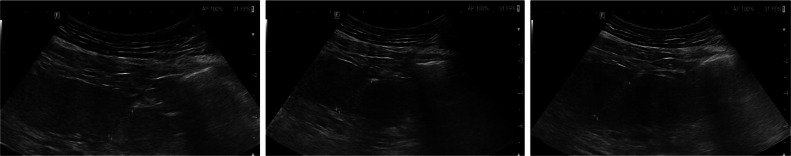
Multinodular steatosis represents a relatively uncommon manifestation of fatty liver disease (FLD). Co-morbidities such as metabolic syndrome or cirrhosis are often associated. Despite typical features of imaging (ultrasound, CT, and MRI), core biopsy sometimes remains the gold standard for diagnosis. We describe the case of a 57-year-old male patient with a long history of hepatic cirrhosis and a recent diagnosis of carcinoma of the tongue, successfully treated. Due to the occurrence of nausea, diarrhea and jaundice the patient is admitted to Our Hospital where ultrasound examination and contrast-enhanced CT are performed, showing global hypoechogenicity of the liver parenchyma with multiple hypo-attenuating lesions. To rule out metastatic lesions, contrast-enhanced CT of the thorax and cranium and gastroscopy and colonoscopy are performed, with no evidence of primary malignancy. Core biopsy reveals macro-vacuolar steatosis within a cirrhotic liver with regenerative aspects.
Keywords: Cirrhosis; Core biopsy; Metastatic disease; Multinodular steatosis.
© 2024 The Authors. Published by Elsevier Inc. on behalf of University of Washington.
Figures

References
-
- Kosobyan EP, Smirnova OM. Current concepts of the pathogenesis of nonalcoholic fatty liver disease. Diabetes Mellitus. 2010;13(1):55–64. doi: 10.14341/2072-0351-6018. - DOI
Publication types
LinkOut - more resources
Full Text Sources
