. 2024 Aug 26;35(1):99-108.
doi: 10.1055/s-0044-1789232. eCollection 2025 Jan.

Radiographs in Pediatric Rheumatology: Where Do We Stand?

Affiliations

Radiographs in Pediatric Rheumatology: Where Do We Stand?

Deeksha Bhalla et al. Indian J Radiol Imaging. .

Abstract

Rheumatic disorders in children include inflammatory arthritis, inflammatory bone disorders such as chronic nonbacterial osteomyelitis (CNO), connective tissue disorders, and vasculitides (juvenile dermatomyositis, scleroderma). The diagnosis in these children is based on a combination of history, clinical examination, and laboratory investigations. Radiographs play an important role in children with arthritis, who have atypical presentation or for assessment of disease-related damage and differentiation from mimics. Further, radiographs also have an ancillary role in the assessment of musculoskeletal disorders such as dermatomyositis and hemophilia. This review seeks to present a detailed analysis of the specific indications and advantages of radiographs in the situations. Further, a structured reporting format for assessment of radiographs in pediatric rheumatic disorders has also been presented for the reader's reference.

Keywords: X-rays; chronic nonbacterial osteomyelitis; juvenile idiopathic arthritis; pediatrics; rheumatology.


Conflict of interest statement

Conflict of Interest None declared.

Figures

Fig. 1
Fig. 1
List the radiographic findings noted in this posteroanterior (PA) radiograph of bilateral wrist and hands. Answer available under the “Reporting Checklist” section.
Fig. 2
Fig. 2
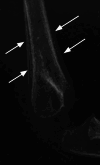
Periosteal reaction in leukemia. Lateral radiograph of the elbow in a 2-year-old boy with pain around the joint shows a lamellated periosteal reaction ( white arrow ).
Fig. 3
Fig. 3
Monoarticular arthritis in an 8-year-old girl. ( A ) On the frontal radiograph, there is symmetric soft-tissue swelling around the right knee joint ( arrow ) suggesting synovial proliferation. ( B ) The lateral view shows synovial proliferation in the infrapatellar bursa, partially effacing the infrapatellar fat pad as well as along the posterior joint recess ( arrows ). Monoarticular knee involvement is a common presentation of oligoarticular juvenile idiopathic arthritis (JIA).
Fig. 4
Fig. 4
Etiological differentiation of dactylitis based on radiographs. ( A ) In a 12-year-old male child patient with juvenile idiopathic arthritis, there is synovial proliferation around the metacarpophalangeal, proximal, and distal interphalangeal joints with periarticular osteopenia ( white arrow ). Note is also made of soft-tissue swelling around the wrist joint and carpal erosions ( black arrow ). ( B ) Bony expansion with lytic areas and periostitis seen in the phalanges and metacarpals in an 18-month-old girl with tubercular dactylitis (spina ventosa).
Fig. 5
Fig. 5
Juvenile spondyloarthropathy in a 16-year-old adolescent male patient. ( A ) There is complete loss of joint space seen in both sacroiliac joints (left > right) along the superior aspect ( arrows ) suggesting bony ankylosis. ( B ) Anteroposterior (AP) radiograph of the pelvis with both hip joints also shows loss of joint space in both the hip joints (right > left) with sclerosis and flattening of the right femoral head ( arrows ).
Fig. 6
Fig. 6
Progressive pseudo-rheumatoid dysplasia in a 9-year-old male child patient with progressive hand deformity and short stature. ( A ) Posteroanterior (PA) radiograph of the hands reveals epiphyseal enlargement of the distal phalanges ( white arrow ) as well as the distal metacarpals ( black arrow ). ( B ) Similar enlargement of metatarsal epiphysis is also seen ( arrow ). ( C ) On lateral spine radiograph, the characteristic anterosuperior end plate defects are noted ( arrow ).
Fig. 7
Fig. 7
Assessment of disease-related damage at the hip joint in a 15-year-old male child patient. ( A ) Frontal radiograph shows pelvic tilt toward the right side. There is loss of joint space with periarticular osteopenia in the right hip joint ( arrow ). ( B ) On the oblique view of the right hip, there is significant joint space narrowing ( arrow ) with flattened femoral head, though no erosion is seen.
Fig. 8
Fig. 8
Assessment of disease-related damage at the wrist joint in a 14-year-old girl. The anteroposterior (AP) radiograph of the right wrist, when scored according to the Ravelli modification of Sharp/van der Heijde method. ( A ) Joint space narrowing (JSN) was assessed at the radio-scaphoid, scapho-trapezoid, scapho-capitate joints, joints between the capitate, hamate and third, fourth, and fifth metacarpal bases, as well as all metacarpophalangeal joints. Additionally, the interphalangeal joints are also assessed (not shown). Black arrows show joints without JSN, while white arrows show joints with JSN. ( B ) Erosions were assessed at the metaphysis of the radius and ulna, scaphoid, lunate, trapezoid, capitate, hamate, bases of the first to fourth metacarpals and proximal phalanges. The bases of distal phalanges are also assessed (not shown). Black arrows depict sites without erosion, while white arrows depict sites with erosion.
Fig. 9
Fig. 9
Cervical spine involvement in juvenile idiopathic arthritis (JIA). ( A ) Lateral radiograph of the cervical spine in a 6-year-old male child patient shows increased atlanto-dental interval ( black arrow ) and reduced posterior atlanto-dental interval (PADI) ( double arrow ). There is apophyseal ankylosis seen in the upper cervical spine ( white arrows ). ( B ) In another patient, a 13-year-old male child patient, the atlanto-dental ( black arrow ) and PADI ( double arrow ) are normal. There is bony ankylosis seen in the vertebral bodies in the upper cervical spine ( white arrow s).
Fig. 10
Fig. 10
Calcinosis cutis in dermatomyositis in a 10-year-old male child patient. ( A ) Linear soft-tissue calcification is seen in both the upper limbs ( arrow ). ( B ) There is more extensive calcification seen around the pelvic bones and in the soft tissue of the medial thigh, forming a sheetlike pattern. ( C ) Nodular deposits are also seen in the soft tissue of both calves.
Fig. 11
Fig. 11
Chronic nonbacterial osteomyelitis in a 9-year-old boy. The posteroanterior (PA) radiograph of the hand shows extensive periostitis ( arrows ) involving the third and fourth metacarpals as well as the distal radius and ulna. Healing lytic sclerotic lesion is seen in the third metacarpal head adjacent to the growth plate ( solid arrow ).
Fig. 12
Fig. 12
Radiograph of bilateral hands and wrist in a 10-year-old boy with joint idiopathic arthritis (JIA). The structured reporting format is shown in Fig. 13 .
Fig. 13
Fig. 13
Structured reporting format for radiographs in juvenile arthritis.
Fig. 14
Fig. 14
Summary of the role of radiography in rheumatic disorders in children.

Similar articles

References

    1. Sheybani E F, Khanna G, White A J, Demertzis J L. Imaging of juvenile idiopathic arthritis: a multimodality approach. Radiographics. 2013;33(05):1253–1273. - PubMed
    1. International League of Associations for Rheumatology . Petty R E, Southwood T R, Manners P et al.International League of Associations for Rheumatology classification of juvenile idiopathic arthritis: second revision, Edmonton, 2001. J Rheumatol. 2004;31(02):390–392. - PubMed
    1. Ravelli A, Martini A. Juvenile idiopathic arthritis. Lancet. 2007;369(9563):767–778. - PubMed
    1. Arkun R, Mete B D. Musculoskeletal brucellosis. Semin Musculoskelet Radiol. 2011;15(05):470–479. - PubMed
    1. Felici E, Novarini C, Magni-Manzoni S et al.Course of joint disease in patients with antinuclear antibody-positive juvenile idiopathic arthritis. J Rheumatol. 2005;32(09):1805–1810. - PubMed

LinkOut - more resources