. 2024 Jul 19;35(1):159-168.
doi: 10.1055/s-0044-1788588. eCollection 2025 Jan.

Sonographic Assessment of Isthmocele and Its Obstetric Complications in Subsequent Pregnancies: A Pictorial Review

Affiliations

Sonographic Assessment of Isthmocele and Its Obstetric Complications in Subsequent Pregnancies: A Pictorial Review

Prateek Agarwal et al. Indian J Radiol Imaging. .

Abstract

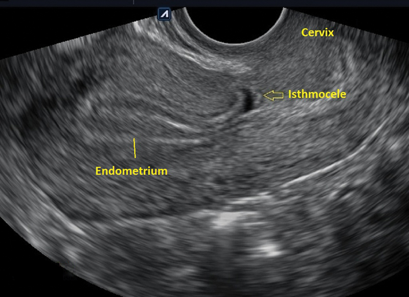
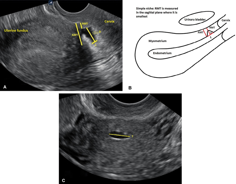
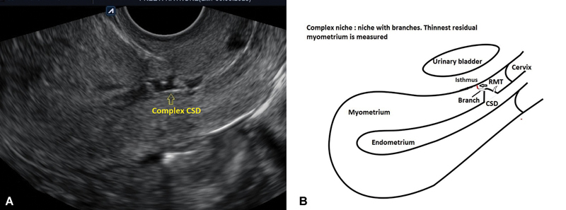
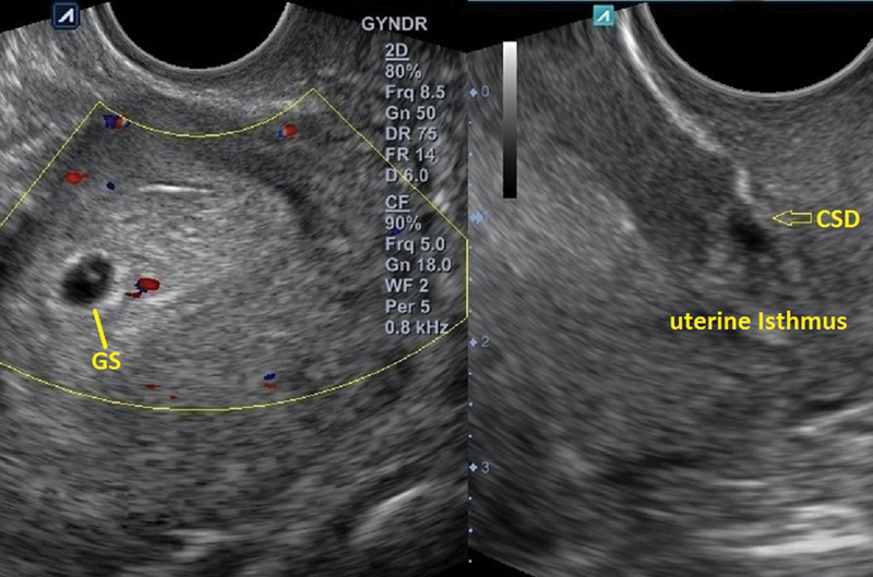
Cesarean scar defect represents a significant pathology attributed to the rising prevalence of cesarean deliveries. While not commonplace, these lesions can give rise to severe obstetric consequences during subsequent pregnancies. Given the potential complications, it is advisable to screen for uterine niches using transvaginal ultrasound (TVUS) or contrast-enhanced TVUS for individuals planning to conceive. Surgical repair and correction of these lesions can be crucial in averting obstetric and perinatal complications in future pregnancies. Furthermore, timely sonographic evaluation and reporting of isthmocele-related obstetric complications can help avoid serious issues.

Keywords: cesarean scar pregnancy; cesarean section; isthmocele; placenta accreta spectrum; retained products of conception; sonography.


Conflict of interest statement

Conflict of Interest None declared.

Figures

Fig. 1
Fig. 1
TVUS in the sagittal plane shows a triangular defect with a base communicating with the endometrial cavity. Assessment of the CSD is recommended after 3 months of a CS, particularly during the mid-follicular phase when fluid is present in the uterine cavity. CS, cesarean section; CSD, cesarean scar defect; TVUS, transvaginal ultrasound.
Fig. 2
Fig. 2
( A,B ) A simple niche in the longitudinal and ( C ) transverse planes: The L, D, and T of a niche are measured in a single plane where it appears largest. The RMT is measured in the sagittal plane where it is the smallest. ( B ) Schematic representation of simple niche. Drawings adapted from Antila-Långsjö et al. AMT, adjacent myometrial thickness; D, depth; L, length; RMT, residual myometrial thickness; T, width.
Fig. 3
Fig. 3
( A, B ) Complex niche (niche with branches): multiple planes might be needed. Length and depth measurements are taken in the same sagittal plane. However, assessing the thinnest RMT of the main niche and its branch might require one or two different sagittal planes. ( B ) Schematic diagram of a complex niche. Uterus line diagram adapted from Antila-Långsjö et al. CSD, cesarean scar defect; RMT, residual myometrial thickness.
Fig. 4
Fig. 4
TVUS reveals a 5-week intrauterine gestational sac with an isthmocele (arrow). The RMT/AMT ratio was <50%. In such cases, the risk of PAS increases if the future placenta is anterior and low lying. The niche can retain POC in the event of spontaneous abortion or if MTP is chosen. AMT, adjacent myometrial thickness; CSD, cesarean scar defect; GS, gestational sac; MTP, medical termination of pregnancy; PAS, placenta accreta spectrum; POC, products of conception; RMT, residual myometrial thickness; TVUS, transvaginal ultrasound.
Fig. 5
Fig. 5
( A, B ) TVUS at 5 weeks of gestation: features of CSP (endogenic type) include an empty uterine cavity, a gestational sac with a yolk sac implanted in the isthmic part of the uterus, a thin anterior myometrium, and an empty cervical canal. ( B ) Diagrammatic representation of endogenic CSP. Uterus line diagram adapted from Antila-Långsjö et al. CS, cesarean section; CSP, cesarean scar pregnancy; RMT, residual myometrial thickness; TVUS, transvaginal ultrasound.
Fig. 6
Fig. 6
TVUS at 7 weeks of gestation: The gestational sac is lying near the CS scar. No thinning of the anterior myometrium; a sliding sign was present suggesting ongoing miscarriage. Previous CS scar (arrows). CS, cesarean section; TVUS, transvaginal ultrasound.
Fig. 7
Fig. 7
( A ) The gestational sac shows a well-formed fetal pole surrounded by a hyperechoic rim of choriodecidual reaction. Note the thinned-out anterior myometrium (arrows). Doppler reveals peripheral vascularity around the gestational sac. ( B ) Note the relation of the gestational sac with the UCL and SL. The major part of the gestational sac is embedded in the myometrium and does not extend beyond the serosal line. [With the kind permission of Dr Amol Karwande] ( C ) Diagrammatic representation of ( B ). Uterus line diagram adapted from Antila-Långsjö et al. CSP, cesarean scar pregnancy; RMT, residual myometrial thickness; SL, serosal line; UCL, uterine cavity line.
Fig. 8
Fig. 8
( A–C ) Sonographic appearances of exogenic type of CSP in retroverted ( A ) and anteverted uterus ( B ). The gestational sac is herniated through the serosa. The exogenic type of CSP is associated with absent residual myometrium and a severe form of PAS due to extensive trophoblastic invasion (with the kind permission of Dr. Nishant Patel). ( C ) Diagrammatic representation of exogenic CSP. Uterus line diagram adapted from Antila-Långsjö et al. CSD, cesarean scar defect; CSP, cesarean scar pregnancy; PAS, placenta accreta spectrum.
Fig. 9
Fig. 9
( A, B ) A known case of CSP diagnosed at 7 weeks of gestation, follow-up ( A ) grayscale and ( B ) color Doppler images at 13 weeks and 5 days show several observations. The absence of myometrium and irregularities in the echogenic bladder line (arrow) is due to abnormal placental vasculature at the ureterovesical interface (indicated by arrows). ( B ) depicts bridging vessels with the color Doppler, matching those seen in ( A ). CSP, cesarean scar pregnancy; UB, urinary bladder.
Fig. 10
Fig. 10
Grayscale and color Doppler images of RPOC at cesarean scar diverticulum. The prior status of CSD was not known. ( A–C ) A 36-year-old woman, with two previous CS, had heavy vaginal bleeding on the 17th day of taking pills for the termination of a 7-week intrauterine viable pregnancy. ( A ) Transabdominal mid-sagittal and transverse grayscale ultrasound revealed a heterogeneous mass in the isthmic part of the uterus protruding through the anterior uterine wall (arrows). ( B ) Grayscale ultrasound performed after 10 days of conservative treatment demonstrated an increase in the size of the mass, resembling a myoma. ( c ) Doppler imaging indicated vascular RPOC. Surgical intervention following failed conservative treatment confirmed RPOC at CSD upon pathology examination. CS, cesarean section; CSD, cesarean scar defect; RPOC, retained products of conception; UB, urinary bladder.
Fig. 11
Fig. 11
Grayscale and color Doppler images of RPOC at cesarean scar diverticulum. The prior status of CSD was not known. ( A, B ) On the 15th day, postsurgical evacuation for a miscarriage involving a 9-week intrauterine pregnancy; ( A ) suprapubic grayscale scan revealed a heterogeneous mass at the previous CS scar. Doppler examination showed no vascularity, indicative of avascular RPOC. However, on ( B ) grayscale and color Doppler TVUS revealed multiple cystic lesions and turbulent flow involving the adjacent myometrium suggestive of EMV. The patient had persistent and uncontrollable vaginal bleeding that did not respond to conservative management or uterine artery embolization. Consequently, a hysterectomy was performed. ( C ) Diagrammatic representation of RPOC at CSD associated with EMV. Uterus line diagram adapted from Antila-Långsjö et al. CS, cesarean section; CSD, cesarean scar defect; EMV, enhanced myometrial vascularity; RPOC, retained products of conception; TVUS, transvaginal ultrasound; UB, urinary bladder.
Fig. 12
Fig. 12
Grayscale and color Doppler images of RPOC at cesarean scar diverticulum. ( A, B ) A suprapubic grayscale and color Doppler scan on the 7th day following termination of ∼12 weeks' gestation. An earlier 7-week scan showed low implantation near the previous CS scar. The patient presented with heavy vaginal bleeding. ( A ) Grayscale scan revealed a heterogeneous mass displaying cystic regions, protruding through the isthmic part of the uterus, the mass exhibited an absence of residual myometrium and alterations at the uterovesical interface (arrows). ( B ) Color Doppler imaging identified abnormal vessels at the uterovesical interface, suggestive of PAS (arrows). Increased vascularity with bidirectional and turbulent flow in the adjacent myometrium suggested EMV (arrows). These findings led to the decision to perform a hysterectomy. CS, cesarean section; CSD, cesarean scar defect; EMV, enhanced myometrial vascularity; PAS, placenta accreta spectrum; POC, products of conception; RPOC, retained products of conception; UB, urinary bladder.
Fig. 13
Fig. 13
Grayscale and color Doppler images of RPOC at cesarean scar diverticulum. ( A, B ) On the 18th day following methotrexate-treated CSP at 6-week gestation, ( A ) suprapubic grayscale and ( B ) color Doppler scans were performed. The patient presented with unmanageable vaginal bleeding. A mass characterized by numerous cystic regions, extending from the CS scar site and protruding through the anterior lower half of the uterus, resembling a molar pregnancy. ( B ) Color Doppler imaging revealed vascular RPOC and EMV. Despite undergoing uterine artery embolization, the bleeding persisted, ultimately leading to a hysterectomy. CS, cesarean section; CSD, cesarean scar defect; CSP, cesarean scar pregnancy; EMV, enhanced myometrial vascularity; POC, products of conception; RPOC, retained products of conception; UB, urinary bladder.
Fig. 14
Fig. 14
TVUS after 10 days of a vaginal delivery; ruptured CS scar (arrows). CS, cesarean section; TVUS, transvaginal ultrasound.

Similar articles

References

    1. van der Voet L F, Bij de Vaate A M, Veersema S, Brölmann H AM, Huirne J AF. Long-term complications of caesarean section. The niche in the scar: a prospective cohort study on niche prevalence and its relation to abnormal uterine bleeding. BJOG. 2014;121(02):236–244. - PubMed
    1. Bij de Vaate A J, Brölmann H AM, van der Voet L F, van der Slikke J W, Veersema S, Huirne J A. Ultrasound evaluation of the Cesarean scar: relation between a niche and postmenstrual spotting. Ultrasound Obstet Gynecol. 2011;37(01):93–99. - PubMed
    1. Glavind J, Madsen L D, Uldbjerg N, Dueholm M. Cesarean section scar measurements in non-pregnant women using three-dimensional ultrasound: a repeatability study. Eur J Obstet Gynecol Reprod Biol. 2016;201:65–69. - PubMed
    1. Fiocchi F, Petrella E, Nocetti L et al.Transvaginal ultrasound assessment of uterine scar after previous caesarean section: comparison with 3T-magnetic resonance diffusion tensor imaging. Radiol Med (Torino) 2015;120(02):228–238. - PubMed
    1. van der Voet L LF, Limperg T, Veersema S et al.Niches after cesarean section in a population seeking hysteroscopic sterilization. Eur J Obstet Gynecol Reprod Biol. 2017;214:104–108. - PubMed

LinkOut - more resources