

Supraspinous Fossa: Anatomy and Pathology
- PMID: 39697518
- PMCID: PMC11651868
- DOI: 10.1055/s-0044-1787667
Supraspinous Fossa: Anatomy and Pathology
Abstract
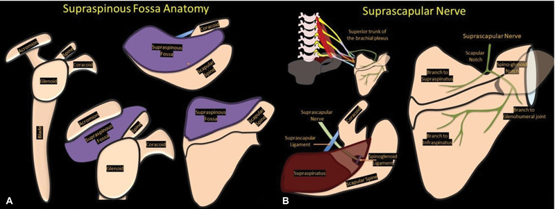
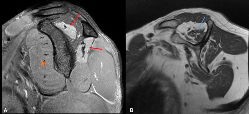
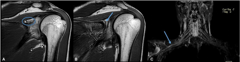
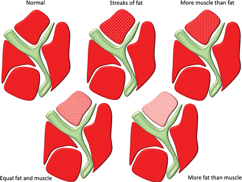
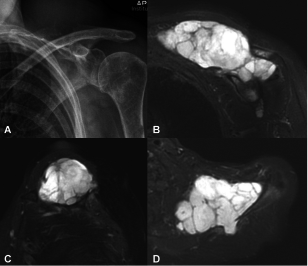
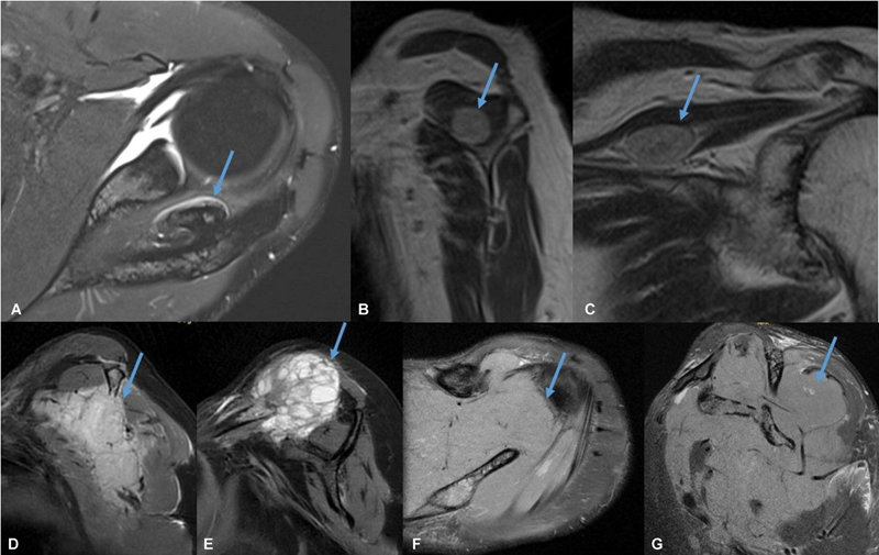
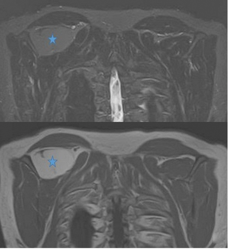
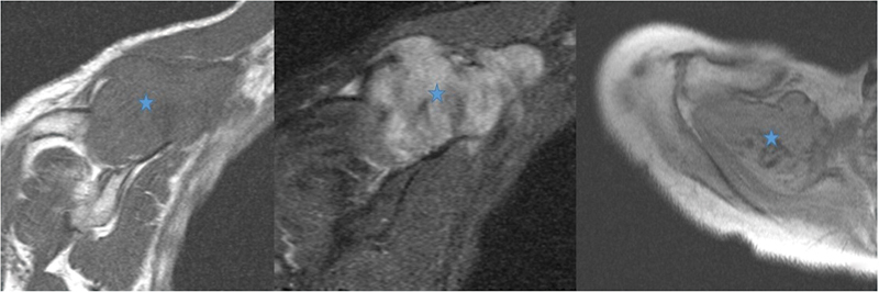
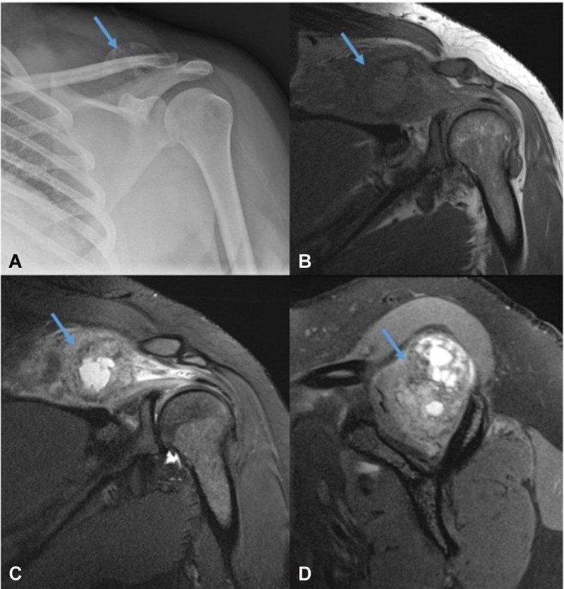
Supraspinous fossa is an important location in the periscapular region, which houses important structures such as the supraspinatus muscle and the suprascapular nerve. The supraspinous fossa can be affected by pathologies involving its contents (supraspinatus muscle and suprascapular nerve), osseous boundary (scapular body, distal clavicle, and spinous process), or superficial soft tissue covering it. In this pictorial review, we describe the detailed anatomy of the supraspinous fossa. We have also covered imaging of wide range of pathologies that can affect supraspinous fossa such as paralabral cyst, muscle edema/atrophy, malignancies (primary and secondary), and miscellaneous lesions (myositis ossificans, fibromatosis, nerve sheath tumor, etc.). An awareness of the imaging findings of these entities is essential for a radiologist to avoid misinterpretation and can aid a timely diagnosis.
Keywords: MRI; suprascapular nerve; supraspinatus muscle; ultrasonography.
Indian Radiological Association. This is an open access article published by Thieme under the terms of the Creative Commons Attribution-NonDerivative-NonCommercial License, permitting copying and reproduction so long as the original work is given appropriate credit. Contents may not be used for commercial purposes, or adapted, remixed, transformed or built upon. ( https://creativecommons.org/licenses/by-nc-nd/4.0/ ).
Conflict of interest statement
Conflict of Interest None declared.
Figures
References
-
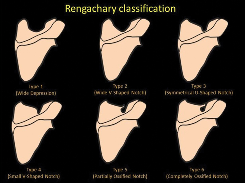
- Rengachary S S, Burr D, Lucas S, Brackett C E. Suprascapular entrapment neuropathy: a clinical, anatomical, and comparative study. Part 3: comparative study. Neurosurgery. 1979;5(04):452–455. - PubMed
-
- Jeno S H, Munjal A, Schindler G S. Treasure Island (FL): StatPearls Publishing; 2022. Anatomy, shoulder and upper limb, arm supraspinatus muscle. - PubMed
-
- Flores D V, Mejía Gómez C, Estrada-Castrillón M, Smitaman E, Pathria M N. MR imaging of muscle trauma: anatomy, biomechanics, pathophysiology, and imaging appearance. Radiographics. 2018;38(01):124–148. - PubMed
Publication types
LinkOut - more resources
Full Text Sources