. 2024 Oct 23;9(1):ziae133.
doi: 10.1093/jbmrpl/ziae133. eCollection 2025 Jan.

Geranylgeranyl diphosphate synthase inhibition impairs osteoclast differentiation, morphology, and resorptive activity

Affiliations

Geranylgeranyl diphosphate synthase inhibition impairs osteoclast differentiation, morphology, and resorptive activity

Molly E Muehlebach et al. JBMR Plus. .

Abstract

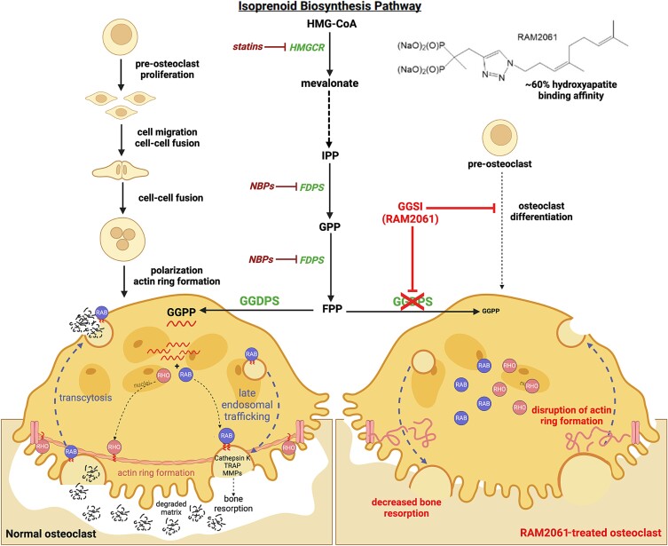
Nitrogen bisphosphonates, such as zoledronic acid, target the enzyme farnesyl diphosphate synthase (FDPS) in the isoprenoid biosynthetic pathway (IBP), and are the frontline treatment for osteolytic bone diseases. A strong affinity of these agents for bone limits their distribution out of the skeleton. Geranylgeranyl diphosphate synthase (GGDPS) is directly downstream to FDPS in the IBP and novel GGDPS inhibitors such as RAM2061 have been shown to have key drug-like features including prolonged half-life, metabolic stability, and systemic distribution. Furthermore, RAM2061 exerts anti-neoplastic benefits in mouse models of multiple myeloma and Ewing sarcoma. Therefore, we are interested in determining the potential impact of RAM2061 on osteoclast biology and bone remodeling. Studies utilizing undifferentiated RAW264.7 cells demonstrated that treatment with RAM2061 depletes cells of geranylgeranyl diphosphate, impairs protein geranylgeranylation, and induces markers of the unfolded protein response pathway and apoptosis. Differentiation of RAW264.7 cells to mature osteoclasts is disrupted by RAM2061, resulting in decreased numbers of mature osteoclasts, altered morphology, and decreased tartrate-resistant acid phosphatase activity. Treatment of fully differentiated RAW264.7 cells with RAM2061 led to decreased resorptive activity. Confocal microscopy studies revealed that RAM2061 disrupts Cdc42 localization, inhibiting proper actin ring formation in osteoclasts. No significant impact on bone turnover markers or bone histomorphology was observed following a 3-week treatment of CD-1 mice with RAM2061, although decreased numbers of osteoclasts were observed. Liquid chromatography-tandem mass spectrometry studies confirmed accumulation of RAM2061 in bone from the in vivo studies as well as hydroxyapatite binding in vitro. In conclusion, these studies are the first to demonstrate the anti-osteoclastic activity of GGDPS inhibitor treatment and support future studies exploring the therapeutic benefit of this novel therapy in the setting of pathological bone remodeling.

Keywords: antiresorptives; molecular pathways—remodeling; osteoclasts; other (therapeutics); preclinical studies.


Conflict of interest statement

The authors have no conflicts of interest.

Figures

Graphical Abstract
Graphical Abstract
Figure 1
Figure 1
RAM2061 demonstrates on-target activity and cytotoxic effects in undifferentiated RAW264.7 cells. (A) RAM2061 and zoledronic acid chemical structures. (B) Levels of intracellular farnesyl pyrophosphate (FPP) and geranylgeranyl diphosphate (GGPP) in RAW264.7 cells following 48 h incubation in the presence or absence of RAM2061 (200 or 400 nM) or zoledronic acid (ZA, 20 μM) (n = 3 biological replicates, data are displayed as mean ± SD; ND = not detected; *** denotes p<.001 per t-test). (C) Immunoblot analysis for unmodified Rap1a (Rap1a) and β-tubulin (loading control) following 24, 48, or 72 h incubation with varying concentrations of RAM2061 in RAW264.7 cells. Immunoblots are representative of 3 independent experiments. (D) MTT assays were performed following a 24, 48, or 72 h incubation period with RAM2061 in RAW264.7 cells (n = 4, data are displayed as mean ± SD).
Figure 2
Figure 2
Geranylgeranyl diphosphate synthase (GGDPS) inhibitor (GGSI) treatment of undifferentiated RAW264.7 cells induces unfolded protein response (UPR) activation and apoptosis. (A) Quantitative real-time polymerase chain reaction analysis of UPR markers ATF4, ATF6, CHOP, IRE1, and PERK following incubation of RAW264.7 cells in the presence or absence of lovastatin (5 μM), zoledronic acid (ZA) (15 μM), or RAM2061 (200 or 400 nM) for 24 or 48 h. Data represent fold-change normalized to control (n = 3 biological replicates, data are represented as mean ± SD; * denotes p<.05, ** denotes p<.01, *** denotes p<.001). (B) Analysis of XBP1 splicing following incubation of RAW264.7 cells in the presence or absence of lovastatin (5 μM), ZA (15 μM), or RAM2061 (200 or 400 nM) for 24 or 48 h. Images are representative of 3 independent experiments. (C) RAW264.7 cells were incubated in the presence or absence of lovastatin, ZA, or RAM2061 for 24 or 48 h. Immunoblot analysis for unmodified Rap1a (Rap1a), UPR markers (ATF4, IRE1, p-e1F2α, e1F2α), apoptotic markers (PARP [arrow = cleaved PARP], cleaved caspases 3, 8, and 9) and β-tubulin (loading control) was performed. Immunoblots are representative of 3 independent experiments. (D) RAW264.7 cells were incubated in the presence or absence of RAM2061 (400 nM) and/or exogenous geranylgeranyl diphosphate (GGPP, 10 μM) for 48 h. Immunoblot analysis for unmodified Rap1a (Rap1a), UPR markers (ATF4, IRE1, p-e1F2α, e1F2α), and apoptotic markers (PARP (arrow = cleaved PARP), cleaved caspases 3, 8, and 9). β-tubulin is shown as the loading control. Immunoblots are representative of 3 independent experiments.
Figure 3
Figure 3
GGSI treatment disrupts osteoclast differentiation. RAW264.7 cells were differentiated over a span of 5 days in the presence or absence of RAM2061 (200 or 400 nM) or ZA (20 μM) (added on day 1 (D1)) (schema in A). On day 5 (D5), cells were fixed and stained with tartrate resistant acid phosphatase (TRAP) to evaluate TRAP-positive osteoclast formation. (A) Images were taken on an EVOS cell imaging microscope at 20x magnification (images are representative of 3 independent experiments; scale bar = 100 μm; arrows indicate multi-nucleated osteoclasts). (B) Total osteoclast number per well was quantified based on TRAP+ staining and the presence of 3 or more nuclei (n = 3 biological replicates, data are represented as mean ± SD, *** denotes p<.001 per t-test). (C) On day 5, media was collected in order to measure levels of secreted TRAP activity (n = 3 biological replicates; data represent secreted TRAP enzymatic activity as percent control ± SD, *** denotes p<.001 per t-test). (D) Quantitative real-time polymerase chain reaction analysis of osteoclast markers ACP5, CTSK, and NFATc1 following incubation in the presence or absence of RAM2061 (200 or 400 nM) or ZA (20 μM) added on day 1 during the differentiation period. On day 5, RNA was extracted and expression was quantified as fold change normalized to control (n = 3 biological replicates, data are represented as mean ± SD; ** denotes p<.01, *** denotes p<.001 per t-test).
Figure 4
Figure 4
GGSI treatment of mature osteoclasts disrupts protein geranylgeranylation and decreases osteoclast number and resorptive function. RAW264.7 cells were differentiated over a span of 5 days and then either RAM2061 or zoledronic acid (ZA) was added on day 5. (A) MTT assays were performed following a 24, 48, or 72 h incubation with RAM2061 (n = 4, data are displayed as mean ± SD). (B) Immunoblot analysis for unmodified Rap1a (Rap1a) and β-tubulin (loading control). -RL = RAW264.7 not stimulated with receptor activator of nuclear factor-κΒ ligand (RANKL); +RL = RAW264.7 differentiated with RANKL. Immunoblots are representative of 3 independent experiments. (C) Schema of differentiation showing timing of RANKL and macrophage colony stimulating factor (MCSF) treatment as well as drug treatment. Following the addition of RAM2061 (200 or 400 nM) or ZA (20 μM) on day 5, cells were fixed on day 7 (D7) and stained with tartrate resistant acid phosphatase (TRAP) to evaluate TRAP-positive osteoclast formation. Images were taken on an EVOS cell imaging microscope at 20x magnification (images are representative of 3 independent experiments; scale bar = 100 μm). (D) Total osteoclast number per well was quantified based on TRAP+ staining and the presence of 3 or more nuclei (n = 3 biological replicates, data are represented as mean ± SD, * denotes p<.05, ** denotes p<.01 per t-test). (E) On day 7, media was collected, and secreted TRAP activity was measured and normalized to control (n = 3 biological replicates; data are displayed as mean ± SD, * denotes p<.05 per t-test). (F) RAW264.7 cells were differentiated over the span of 5 days on hydroxyapatite-coated plates with the subsequent addition of RAM2061 (200 or 400 nM) or ZA (20 μM) on day 5. On day 7, cells were removed and the resorptive area was quantified using ImageLab. Representative images of the resorptive areas taken on an EVOS cell imaging microscope at 20x magnification) (n = 4 biological replicates, data represented as mean ± SD, * denotes p<.05, ** denotes p<.01 per t-test). The arrows indicate resorbed areas; * indicate pits where the entire hydroxyapatite coating was resorbed.
Figure 5
Figure 5
GGSI treatment disrupts Cdc42 GTPase localization and subsequent regulation of actin dynamics necessary for osteoclast differentiation and actin ring formation. RAW264.7 cells were differentiated on glass coverslips over a span of 5 days and RAM2061 (200 or 400 nM) or zoledronic acid (ZA) (20 μM) was added on day 1. (A) On day 5 (D5), osteoclasts were fixed and stained with an antibody against Cdc42 and DAPI (nucleus label) or (B) stained with phalloidin for identification of actin and DAPI (nuclei). (C) Fully differentiated D5 osteoclasts were treated with RAM2061 (200 or 400 nM) or ZA (20 μM) for 48 h. On day 7 (D7), osteoclasts were fixed and stained with an antibody against Cdc42 and DAPI (nucleus label) or (D) stained with phalloidin (actin) and DAPI (nuclei). Images were obtained using a Zeiss 710 LSM at 40x magnification. Images are representative of 3 independent experiments (scale bar = 20 μm).
Figure 6
Figure 6
RAM2061 does not significantly impact normal bone remodeling in CD-1 mice. (A) Schematic representation of drug administration/dosing and downstream readouts conducted. Abbreviations: procollagen type 1 N-propeptide (P1NP); C-terminal telopeptide (CTX), tartrate resistant acid phosphatase 5b (TRAP5b). (B) Immunoblot analysis of unmodified Rap1a and GAPDH (loading control) using 30 μL of bone homogenate from long bones of mice; positive control = cell lysate from RAW264.7 osteoclasts (D5) treated with 400 nM RAM2061 for 48 h). (C) Quantification of RAM2061 accumulation in long bones and mandibles of CD-1 mice (n = 5 per group, data are represented as mean ± SD). (D) MicroCT analysis portraying morphometric analysis of the proximal tibia specimens. Individual data points are shown with mean ± SD (PBS n = 5; RAM2061 n = 5; zoledronic acid (ZA) n = 4; * denotes p<.05, ** denotes p<.01 per t-test). (E) The growth plate in the distal femur of tartrate resistant acid phosphatase (TRAP)-stained sections were identified and 1000 μm from the most distal area was used to encapsulate the area of interest. Quantification of osteoclast numbers in the defined area per femur was performed. Individual data points are shown with mean ± SD (PBS n = 5; RAM2061 n = 4; ZA n = 4, * denotes p<.05, *** denotes p<.001 per t-test). (F) Representative images of TRAP-stained sections of distal femurs (scale bar = 50 μm). The arrows indicate TRAP+ osteoclasts (Tb = trabecular bone, BM = bone marrow).

Similar articles

Cited by

References

    1. Roberts PJ, Mitin N, Keller PJ, et al. . Rho family GTPase modification and dependence on CAAX motif-signaled posttranslational modification. J Biol Chem. 2008;283(37):25150-25163. 10.1074/jbc.M800882200 - DOI - PMC - PubMed
    1. Gomes AQ, Ali BR, Ramalho JS, et al. . Membrane targeting of Rab GTPases is influenced by the prenylation motif. Mol Biol Cell. 2003;14(5):1882-1899. 10.1091/mbc.e02-10-0639 - DOI - PMC - PubMed
    1. Palsuledesai CC, Distefano MD. Protein prenylation: enzymes, therapeutics, and biotechnology applications. ACS Chem Biol. 2015;10(1):51-62. 10.1021/cb500791f - DOI - PMC - PubMed
    1. Yu Z, Surface LE, Park CY, et al. . Identification of a transporter complex responsible for the cytosolic entry of nitrogen-containing bisphosphonates. eLife. 2018;7:e36620. 10.7554/eLife.36620 - DOI - PMC - PubMed
    1. van Beek E, Pieterman E, Cohen L, Löwik C, Papapoulos S. Farnesyl pyrophosphate synthase is the molecular target of nitrogen-containing bisphosphonates. Biochem Biophys Res Commun. 1999;264(1):108-111. 10.1006/bbrc.1999.1499 - DOI - PubMed

LinkOut - more resources