Review
. 2024 Jul 5;5(3):344-357.
doi: 10.20517/evcna.2024.19. eCollection 2024.

Endosomal escape mechanisms of extracellular vesicle-based drug carriers: lessons for lipid nanoparticle design

Affiliations
Review

Endosomal escape mechanisms of extracellular vesicle-based drug carriers: lessons for lipid nanoparticle design

Lasse Hagedorn et al. Extracell Vesicles Circ Nucl Acids. .

Abstract

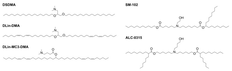
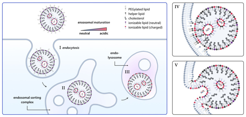
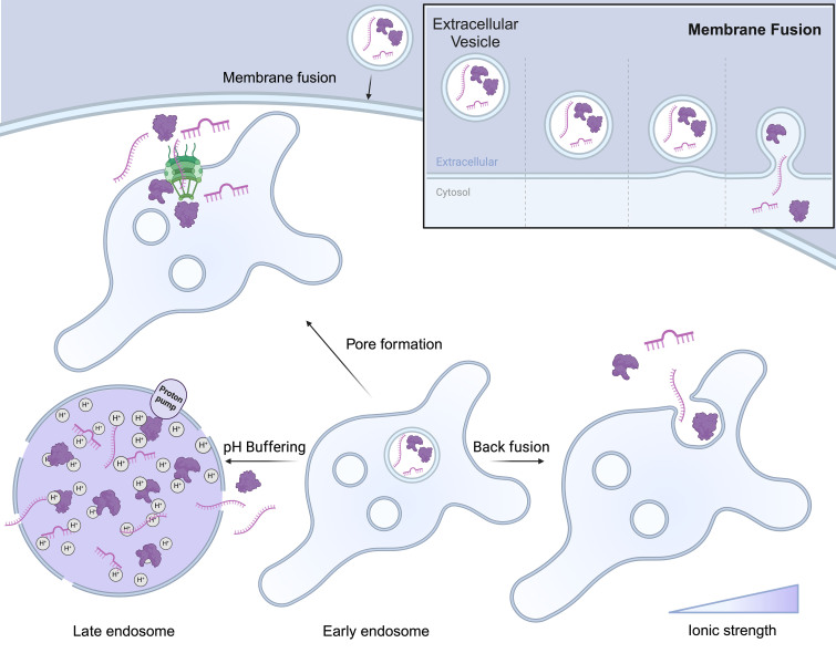
The rise of biologics and RNA-based therapies challenges the limitations of traditional drug treatments. However, these potent new classes of therapeutics require effective delivery systems to reach their full potential. Lipid nanoparticles (LNPs) have emerged as a promising solution for RNA delivery, but endosomal entrapment remains a critical barrier. In contrast, natural extracellular vesicles (EVs) possess innate mechanisms to overcome endosomal degradation, demonstrating superior endosomal escape (EE) compared to conventional LNPs. This mini review explores the challenges of EE for lipid nanoparticle-based drug delivery, and offers insights into EV escape mechanisms to advance LNP design for RNA therapeutics. We compare the natural EE strategies of EVs with those used in LNPs and highlight contemporary LNP design approaches. By understanding the mechanisms of EE, we will be able to develop more effective drug delivery vehicles, enhancing the delivery and efficacy of RNA-based therapies.

Keywords: RNA therapeutics; drug delivery; endosomal escape; extracellular vesicles; lipid nanoparticles; membrane fusion.


Conflict of interest statement

Merkel OM is a consultant for Corden Pharma GmbH, AMW GmbH, and PARI Pharma GmbH. Furthermore, Merkel OM is an advisory board member for Coriolis Pharma GmbH and a consultant for AbbVie Deutschland GmbH on unrelated projects. The other authors have declared that there are no conflicts of interest.

Figures

Figure 1
Figure 1
Ionizable lipids used in LNP formulations. Ionizable lipids used in clinical trials mostly contain head groups with pKa values below 7. Furthermore, the ratio between head group and tail group volume has been optimized to reach the optimal lipid per membrane area for sufficient endosomal escape. This resulted in different lipids with 2-4 alkyl tails with different degrees of saturation of the alkyl tails. LNP: Lipid nanoparticle.
Figure 2
Figure 2
Endosomal escape of LNPs. Following endocytosis, LNPs follow the endo-lysosomal pathway. With the environment becoming increasingly acidic, ionizable lipids become protonated again. Now, the positively charged ionizable lipids can interact with negative membrane lipids to facilitate fusion with the endosomal membrane and release of the nucleic acid into the cytosol. Parts of this figure were created using Biorender. LNPs: Lipid nanoparticles.
Figure 3
Figure 3
Escape mechanisms of extracellular vesicles. There are four proposed mechanisms for delivering EV cargo into the cytoplasm. The first mechanism involves the direct fusion of the EV with the cellular membrane, resulting in the complete release of the EV’s contents. The second mechanism, known as back fusion, occurs when the EV fuses with the endosomal membrane after being taken up via endocytosis. In the third mechanism, following endosomal uptake, EVs can induce pore formation, allowing the cargo to be transported into the cytoplasm. Finally, EVs can promote endosomal escape by buffering acidification and increasing the number of protons transported into the endosome, leading to membrane leakage through osmotic pressure caused by high ionic strength. This figure was created using Biorender. EV: Extracellular vesicle.

Similar articles

Cited by

References

    1. Roberts TC, Langer R, Wood MJA. Advances in oligonucleotide drug delivery. Nat Rev Drug Discov. 2020;19:673–94. doi: 10.1038/s41573-020-0075-7. - DOI - PMC - PubMed
    1. Damase TR, Sukhovershin R, Boada C, Taraballi F, Pettigrew RI, Cooke JP. The limitless future of RNA therapeutics. Front Bioeng Biotechnol. 2021;9:628137. doi: 10.3389/fbioe.2021.628137. - DOI - PMC - PubMed
    1. Dowdy SF. Overcoming cellular barriers for RNA therapeutics. Nat Biotechnol. 2017;35:222–9. doi: 10.1038/nbt.3802. - DOI - PubMed
    1. Mitchell MJ, Billingsley MM, Haley RM, Wechsler ME, Peppas NA, Langer R. Engineering precision nanoparticles for drug delivery. Nat Rev Drug Discov. 2021;20:101–24. doi: 10.1038/s41573-020-0090-8. - DOI - PMC - PubMed
    1. Kesharwani P, Banerjee S, Gupta U, et al. PAMAM dendrimers as promising nanocarriers for RNAi therapeutics. Mater Today. 2015;18:565–72. doi: 10.1016/j.mattod.2015.06.003. - DOI

LinkOut - more resources