

Topical insulin used alone or in combination with drug-depository contact lens for refractory cases of neurotrophic keratopathy
- PMID: 39697672
- PMCID: PMC11652743
- DOI: 10.1016/j.ajoc.2024.102227
Topical insulin used alone or in combination with drug-depository contact lens for refractory cases of neurotrophic keratopathy
Abstract
Purpose: To report the clinical outcomes achieved in refractory cases of neurotrophic keratopathy (NK) through the utilization of insulin eye drops alone or in conjunction with a drug-depository contact lens (DDCL).
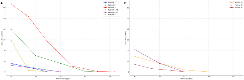
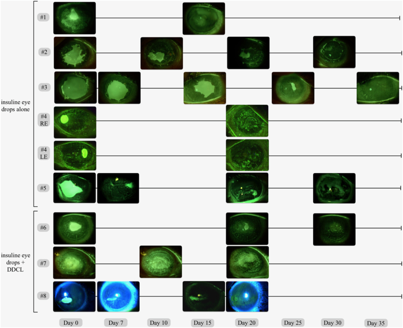
Observations: This multicentric prospective open-label uncontrolled case series included consecutive patients with NK refractory to conventional treatment. Insulin eye drops (1 unit/mL) were prescribed 4 times/day in all cases, and a Therapeutic Hyper-CL™ soft contact lens (EyeYon Medical, Ness Ziona, Israel), designed to act as a drug reservoir, was applied in selected patients. Data about stage and duration of NK, corneal sensitivity, previous treatments, rate and speed of healing, changes of NK area over time were collected. Nine eyes of 8 patients (mean age 52.50 ± 12.03 years [95 % CI, 44.13-60.87]) affected by NK refractory to conventional medical therapy were included. All patients received topical insulin, while DDCL was also applied in 3 cases. At T0, the mean area of the corneal epithelial defect was 21.84 ± 18.35 mm2 [95 % CI, 9.86-33.84]. Complete corneal re-epithelialization occurred in all cases, after a mean time interval of 25.78 ± 8.39 days [95 % CI, 20.30-31.26]. Mean reduction rate of epithelial defect areas was -0.81 ± 0.44 mm2/day [95 % CI, -1.16 to -0.46] for patients treated with insulin eye drops, and -0.63 ± 0.30 mm2/day [95 % CI, -0.96 to -0.29] for those treated with insulin eye drops plus DDCL (p = 0.71). Neither adverse events nor episodes of NK recurrence were reported.
Conclusions and importance: Topical insulin, used alone or in combination with DDCL, is an accessible, inexpensive, and effective treatment for refractory NK.
Keywords: Hyper-CL; Insulin eye drops; NK; Neurotrophic keratopathy; Persistent epithelial defect.
© 2024 The Authors.
Conflict of interest statement
The authors declare that they have no known competing financial interests or personal relationships that could have appeared to influence the work reported in this paper.
Figures
References
Publication types
LinkOut - more resources
Full Text Sources
