

A case documenting distinct natural history of multizonal outer retinopathy and retinal pigment epitheliopathy (MORR) with longitudinal multi-modal documentation of progression
- PMID: 39697673
- PMCID: PMC11653137
- DOI: 10.1016/j.ajoc.2024.102222
A case documenting distinct natural history of multizonal outer retinopathy and retinal pigment epitheliopathy (MORR) with longitudinal multi-modal documentation of progression
Abstract
Purpose: To describe the clinical and imaging characteristics of the acute progressive phase of a recently proposed clinical entity, Multizonal Outer Retinopathy and Retinal Pigment Epitheliopathy (MORR), a variant of Acute Zonal Occult Outer Retinopathy (AZOOR).
Methods: Single observational case report.
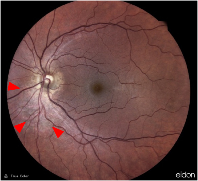
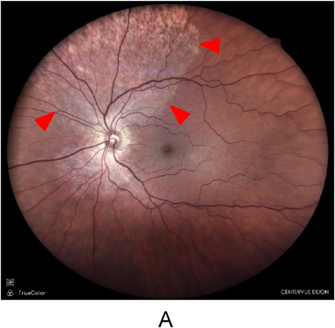
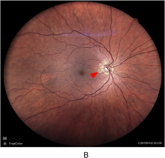
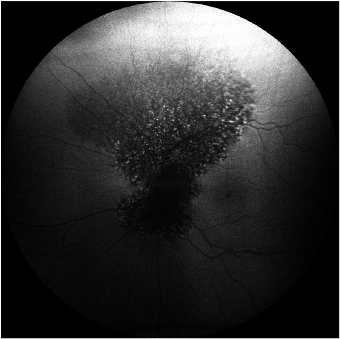
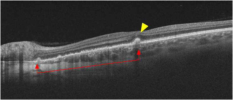
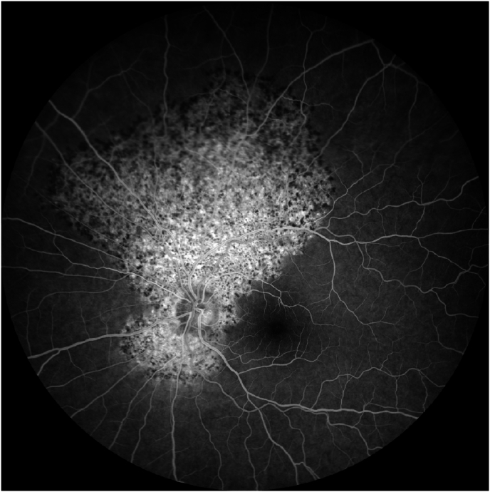
Results: We present the case of a 49-year-old myopic female with progressive outer retinopathy most consistent with a diagnosis of MORR. Through multimodal imaging and longitudinal follow-up, we delineate the clinical course and imaging findings of asymmetrical episodic progressive centrifugal extension of retinal pigment epithelial disturbance in both eyes, highlighting the features of an acute progressive episode not previously described.
Conclusions: Clinicians should be aware of the active clinical and multimodal imaging features of MORR and its distinction from other outer retinopathies due to its sight-threatening distinct clinical course, bilateral involvement with peripapillary lesions, and episodic progression into the macula. Additionally, we describe a "grass-fire" and "spot-fire" progression pattern during acute exacerbation, highlighting the need for vigilant monitoring and early intervention in MORR.
Keywords: AAOR; AZOOR; MORR; Outer retinopathy; White dot syndromes.
Crown Copyright © 2024 Published by Elsevier Inc.
Conflict of interest statement
The authors declare the following financial interests/personal relationships which may be considered as potential competing interests: The authors have no conflict of interest.
Figures

Similar articles
-
MULTIZONAL OUTER RETINOPATHY AND RETINAL PIGMENT EPITHELIOPATHY (MORR): A Newly Recognized Entity or an Unusual Variant of AZOOR?Retina. 2023 Nov 1;43(11):1890-1903. doi: 10.1097/IAE.0000000000003927. Epub 2023 Oct 19. Retina. 2023. PMID: 37748093 Free PMC article.
-
Multizonal outer retinopathy and retinal pigment epitheliopathy (MORR) with a chronologically divergent presentation- a case report.J Ophthalmic Inflamm Infect. 2025 Jul 22;15(1):57. doi: 10.1186/s12348-025-00519-0. J Ophthalmic Inflamm Infect. 2025. PMID: 40694285 Free PMC article.
-
Current understanding of acute zonal occult outer retinopathy (AZOOR).Indian J Ophthalmol. 2024 Jul 1;72(7):935-937. doi: 10.4103/IJO.IJO_3228_23. Epub 2024 Mar 8. Indian J Ophthalmol. 2024. PMID: 38454854 Free PMC article. Review.
-
Acute zonal occult outer retinopathy: a classification based on multimodal imaging.JAMA Ophthalmol. 2014 Sep;132(9):1089-98. doi: 10.1001/jamaophthalmol.2014.1683. JAMA Ophthalmol. 2014. PMID: 24945598
-
Syphilitic Outer Retinopathy: A Case Report and Review of the Literature.J Vitreoretin Dis. 2021 Jun 21;6(1):63-70. doi: 10.1177/24741264211018300. eCollection 2022 Jan-Feb. J Vitreoretin Dis. 2021. PMID: 37007722 Free PMC article. Review.
References
-
- Gass J.D., Agarwal A., Scott I.U. Acute zonal occult outer retinopathy: a long-term follow-up study. Am J Ophthalmol. 2002;134:329–339. - PubMed
-
- Mrejen S., Khan S., Gallego-Pinazo R., Jampol L., Yannuzzi L. Acute zonal occult outer retinopathy A classification based on multimodal imaging. JAMA Ophthalmol. 2014;132(9):1089–1098. - PubMed
LinkOut - more resources
Full Text Sources
