. 2024 Nov 6;10(1):veae093.
doi: 10.1093/ve/veae093. eCollection 2024.

Identification of a novel mycovirus belonging to the "flexivirus"-related family with icosahedral virion

Affiliations

Identification of a novel mycovirus belonging to the "flexivirus"-related family with icosahedral virion

Chien-Fu Wu et al. Virus Evol. .

Abstract

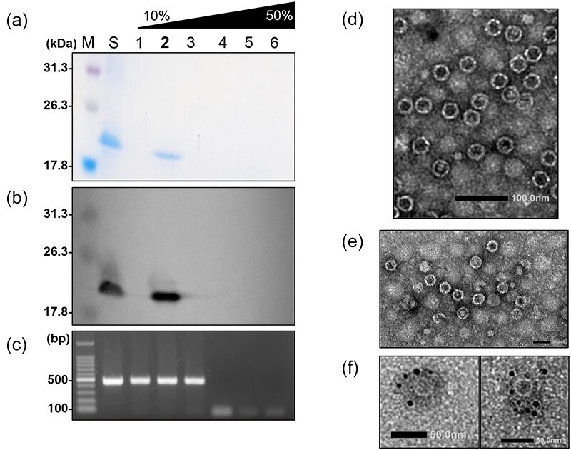
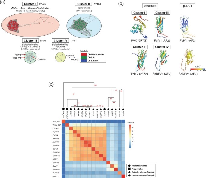
The order Tymovirales currently comprises five viral families with positive-sense RNA [(+)RNA] genomes that infect plants, fungi, and insects. Virion morphologies within the order Tymovirales differ between families, with icosahedral virions in the Tymoviridae and filamentous virions in the other "flexi"viridae families. Despite their different morphologies, these viruses are placed in the same order based on phylogenetic analyses of replicase-associated polyproteins. However, one of the families in the Tymovirales, Deltaflexiviridae, is considered to be capsidless because there have been no published reports of virion isolation. Here, we report that a new "flexivirus"-related (+)RNA virus, prospectively named Fusarium oxysporum icosahedral virus 1 (FoIV1), is icosahedral and that most deltaflexiviruses may have icosahedral virions. Phylogenetic analyses based on replicase-associated polyproteins indicated that FoIV1 forms a distinct group in the Tymovirales with some viruses originally assigned to the Deltaflexiviridae. Electron microscopy, protein analysis, and protein structure predictions indicate that FoIV1 open reading frame 4 encodes a single jelly-roll (SJR)-like coat protein (CP) that constitutes the icosahedral virions. Results of clustering analyses based on amino acid sequences and predicted CP structures suggested that most of the deltaflexiviruses have icosahedral virions composed of SJR-like CPs as in FoIV1, rather than having filamentous virions or capsidless. These results challenge the conventional understanding of viruses in the order Tymovirales, with important implications for revising its taxonomic framework and providing insights into the evolutionary relationships within this diverse and broad host range group of (+)RNA viruses.

Keywords: Tymovirales; coat protein; deltaflexivirus; mycovirus; single jelly-roll; virus particle.


Conflict of interest statement

None declared.

Figures

Figure 1.
Figure 1.
Novel RNA virus in Fusarium oxysporum f. sp. melonis. (a) Colony morphology of F. oxysporum f. sp. melonis strain Fom 405 cultured on PDA for 5 days at 25°C. (b) dsRNA isolated from F. oxysporum f. sp. melonis strain Fom 405 was analyzed on a 1% agarose gel with EtBr (0.5 μg/ml) at 18 V for 20 h. Lane M: 250 ng of λ-EcoT14I-digested DNA marker. (c) Schematic genome organization of FoIV1. Nucleotide sequence of the 5′ terminus of hypothetical sgRNA is shown according to the 5′-RLM-RACE result (Supplementary Fig. S3). ORFs are shown as boxes, ORF1, replicase-associated protein. Mtr, methyl transferase; Hel, helicase; RdRp, RNA-dependent RNA polymerase. ORF2, ORF3, ORF4, and ORF5 are hypothetical proteins.
Figure 2.
Figure 2.
Phylogenetic analyses of FoIV1 and viruses in the order Tymovirales using the aa sequences of replicase-associated proteins. (a) An ML tree was constructed using IQ-TREE v1.6.12. The consensus tree was obtained from 1000 replicates with ultrafast bootstrap analysis (best-fit model: LG + F + R6). Bootstrap values (%) are indicated next to the nodes. (b) A BI tree was constructed using MrBayes 3.2.7 with 100,000 generations. Posterior probabilities are next to the nodes. (c) An NJ tree was constructed using MEGA11. The consensus tree was obtained from 1000 replicates. Bootstrap values (%) are marked next to the branches. Virus marked with “*” was discovered from metagenomic data of plant tissues with potential fungal infection. Virus marked with “**” was detected from plant tissues and sequence was determined using Sanger sequencing. (d) A pairwise identity matrix was generated using SDT v1.3. The numbers shown in the matrix are the pairwise identity (%). The RdRp sequence of FoIV1 identified in this study is highlighted in red. (e) Multiple alignment of RdRp aa sequences of FoIV1, FoIV1-related viruses, and deltaflexiviruses, visualized using pyMSAviz v0.4.2.
Figure 2.
Figure 2.
Phylogenetic analyses of FoIV1 and viruses in the order Tymovirales using the aa sequences of replicase-associated proteins. (a) An ML tree was constructed using IQ-TREE v1.6.12. The consensus tree was obtained from 1000 replicates with ultrafast bootstrap analysis (best-fit model: LG + F + R6). Bootstrap values (%) are indicated next to the nodes. (b) A BI tree was constructed using MrBayes 3.2.7 with 100,000 generations. Posterior probabilities are next to the nodes. (c) An NJ tree was constructed using MEGA11. The consensus tree was obtained from 1000 replicates. Bootstrap values (%) are marked next to the branches. Virus marked with “*” was discovered from metagenomic data of plant tissues with potential fungal infection. Virus marked with “**” was detected from plant tissues and sequence was determined using Sanger sequencing. (d) A pairwise identity matrix was generated using SDT v1.3. The numbers shown in the matrix are the pairwise identity (%). The RdRp sequence of FoIV1 identified in this study is highlighted in red. (e) Multiple alignment of RdRp aa sequences of FoIV1, FoIV1-related viruses, and deltaflexiviruses, visualized using pyMSAviz v0.4.2.
Figure 3.
Figure 3.
FoIV1 virus purification. Purified virus suspension was resolved with a 10–50% CsCl gradient and then fractioned into six fractions. Lane S, virus suspension before resolved in a CsCl gradient. (a) SDS-PAGE electrophoresis of the CsCl gradient fractions. The fractions were resolved in a 13% polyacrylamide gel at 120 V for 2.5 h and then stained with CBB. (b) Western blot analysis of CsCl gradient fractions. Protein bands were detected with antiserum raised against FoIV1 ORF4-encoded protein. (c) Agarose gel electrophoresis of RT-PCR products using the primer pair ORF4-F and ORF4-R (Supplementary Table S2) against full-length FoIV1 ORF4. Fraction 2 showed an 18.2 kDa viral protein and viral nucleic acids and then was subjected to (d) conventional TEM, scale bar: 100 nm. (e) ISEM, scale bar: 60 nm. (f) IEM. Size of the gold particles: 5 nm. Scale bar: 50 nm. Virus particles are ∼32 nm in diameter (buoyant density: 1.26 g/cm3).
Figure 4.
Figure 4.
Sequence- and structure-based similarity analyses of the CPs of FoIV1, FoIV1-related viruses, and other members of the Tymovirales. (a) Sequence-based clustering of CPs of the Tymovirales. CP sequences were clustered using all vs. all pairwise alignment of DIAMOND v2.0.15 (E-value ≤0.001) and then visualized by Cytoscape v3.10.1. Four distinct clusters and two independent nodes are present. Numbers of nodes (n) are marked under the clusters. Nodes are colored by their protein folding types. The red arrows indicate the CPs of FoIV1-related viruses. (b) Protein structure modeling of CPs. Structures are color-coded with a rainbow color scheme from N-terminal (blue) to C-terminal (red). The predicted local distance difference test (pLDDT) scores of Alphafold2 (AF2)-predicted proteins are also shown in the figure. BIDG-CHEF β-strands of SJR folding structures are annotated on the model of TYMV CP. (c) Dendrogram and heatmap of all-against-all structure comparison of SJR-CP of TYMV, Phlebo NC-like CP of PVX, and CPs of FoIV1-related viruses and deltaflexiviruses. The “average” method was used for clustering (clustering methods: ward.D, ward.D2, single, complete, average, mcquitty, median, and centroid were tested to find the best clustering). Bootstrap values are indicated as red numbers at the nodes.

Similar articles

Cited by

References

    1. Adams MJ, Candresse T, Hammond J et al. Family—Betaflexiviridae. In: King AMQ, Adams MJ, Carstens EB et al. (eds), Virus Taxonomy, 9th edn. San Diego: Elsevier, 2012a, 920–41.
    1. Adams MJ, Kreuze JF, Martelli GP. Order—Tymovirales. In: King AMQ, Adams MJ, Carstens EB et al. (eds), Virus Taxonomy, 9th edn. San Diego: Elsevier, 2012b, 901–03.
    1. Andika IB, Wei S, Cao C et al. Phytopathogenic fungus hosts a plant virus: a naturally occurring cross-kingdom viral infection. Proc Natl Acad Sci U S A 2017;114:12267–72. doi: 10.1073/pnas.1714916114 - DOI - PMC - PubMed
    1. Barrio-Hernandez I, Yeo J, Jänes J et al. Clustering predicted structures at the scale of the known protein universe. Nature 2023;622:637–45. doi: 10.1038/s41586-023-06510-w - DOI - PMC - PubMed
    1. Bartholomäus A, Wibberg D, Winkler A et al. Deep sequencing analysis reveals the mycoviral diversity of the virome of an avirulent isolate of Rhizoctonia solani AG-2-2 IV. PLoS One 2016;11:e0165965. doi: 10.1371/journal.pone.0165965 - DOI - PMC - PubMed

LinkOut - more resources