. 2024 Dec 18;17(12):2221-2228.
doi: 10.18240/ijo.2024.12.09. eCollection 2024.

Treatment of superficial corneal opacities with corneal stromal lenticule obtained through SMILE surgery

Affiliations

Treatment of superficial corneal opacities with corneal stromal lenticule obtained through SMILE surgery

Shi-Si Hu et al. Int J Ophthalmol. .

Abstract

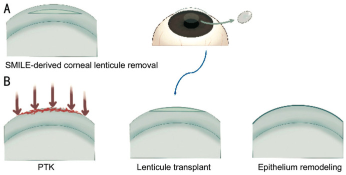
Aim: To evaluate the clinical efficacy and feasibility of superficial corneal opacities treated by excimer laser phototherapeutic keratectomy (PTK) combined with small incision lenticule extraction (SMILE)-derived corneal stromal lenticule transplantation.

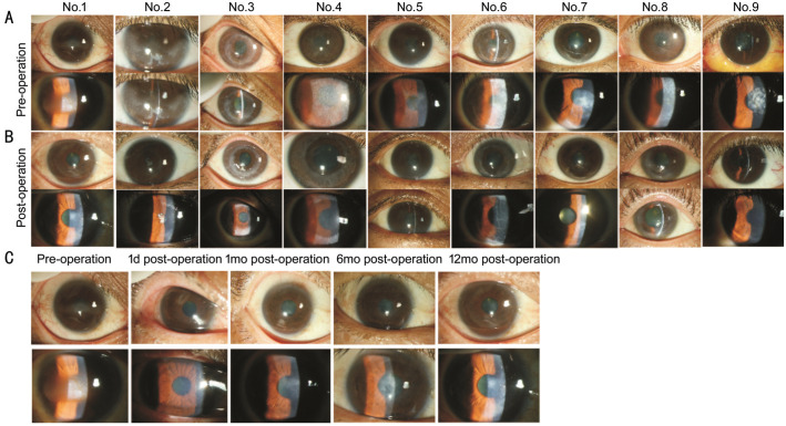
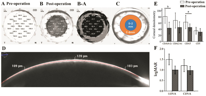
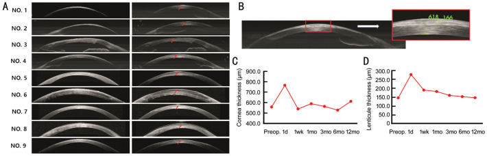
Methods: A retrospective interventional case series of nine patients aged 12-59y with superficial corneal opacity caused by different pathologies who underwent standardized PTK combined with SMILE-derived corneal stromal lenticule transplantation was examined. Lenticule patches were fixed with fibrin glue. All patients underwent pre- and post-operative clinical assessments at different times for up to 12mo. Slit lamp microscopy, corneal density, uncorrected distance visual acuity (UDVA), corrected distance visual acuity (CDVA), and anterior segment optical coherence tomography (AS-OCT) were examined.

Results: The patients' mean age was 36.00±5.80 (12-59)y. Seven eyes (77.8%) gained UDVA and CDVA at the last measurement compared to their preoperative levels. The densities of the total cornea, the total anterior corneal layer, and the anterior corneal layers of 0-2 and 2-6 mm decreased significantly by 12.4%, 27.5%, 46.7%, and 32.8%, respectively. After human allogeneic transplantation, the implanted lenticules of all eyes were clearly visible by AS-OCT and remained transparent without displacement or graft rejection. The thickness of the central cornea and corneal lenticule transplants were stable throughout the entire postoperative period. One case experienced the postoperative complication of delayed corneal epithelial healing.

Conclusion: PTK combined with SMILE-derived corneal lenticule transplantation improves long-term visual acuity. Therefore, it is a new, safe, and effective method for treating superficial corneal opacity.

Keywords: corneal lenticule; corneal opacity; phototherapeutic keratectomy; transplant.


Conflict of interest statement

Conflicts of Interest: Hu SS, None; Ding H, None; Meng XY, None; Ouyang BW, None; Yang ZD, None; Chen XD, None; Zhong XW, None.

Figures

Figure 1
Figure 1. Graphical representation of PTK combined with SMILE-derived corneal stromal lenticule transplantation procedure
A: The corneal lenticule acquisition procedure during SMILE surgery; B: The PTK combined with epikeratophakia procedure. PTK: Phototherapeutic keratectomy; SMILE: Small incision lenticule extraction.
Figure 2
Figure 2. Pre- and post-operative anterior photographs for patients 1–9 obtained via slit-lamp microscopy
A: The preoperative slit-lamp microscopy images of the nine patients showed banded or localized dense corneal opacity in patients 1, 2, 5, 7, and 9 and superficial granular and confluent deposits with diffuse stromal haze in patients 3, 4, 6, and 8; B: The slit-lamp microscopy images of the same patients at their final postoperative visit demonstrate how corneal opacities can be successfully removed from the optical zone of the cornea without corneal ulceration or neovascularization; C: The postoperative slit-lamp microscopy images of patient 1. The corneal tissue maintained its transparency over time with no visible tissue boundary.
Figure 3
Figure 3. Pre and post-operative corneal densitometry measurements for patients 1–9
A: Preoperative corneal densitometry shows high corneal densities in the area of opacity; B: Postoperative corneal densitometry measurements show decreasing corneal densities in the operated area; B-A: Pre- to post-operative corneal densitometry differences; C: The cornea was divided into four regions based on its diameter: 0–2, 2–6, 6–10, and 10–12 mm; D: A cross-section of the cornea. The red line delineates the most anterior 120 µm of the cornea; E: Comparison of pre- and post-operative (final visit) corneal densitometry for all nine operated eyes. Density decreased significantly in the 0–2 mm region of the superficial cornea and the entire superficial cornea. It also decreased in the 2–6 mm region of the superficial cornea and the entire cornea. Data were presented as the mean±SEM. aP<0.05; bP<0.01. F: Comparison of pre- and post-operative (final visit) UDVA and CDVA for all nine operated eyes. Data were presented as the mean±SEM. UDVA: Uncorrected distance visual acuity; CDVA: Corrected distance visual acuity; SEM: Standard error of the mean; CDA (0–2): The density of the anterior corneal layers of 0–2 mm; CDA (2–6): The density of the anterior corneal layers of 2–6 mm; CDAT: The density of the total anterior corneal layer; CDT: The density of the total cornea.
Figure 4
Figure 4. Representative corneal AS-OCT images
A: Pre- and post-operative corneal AS-OCT images. The red arrowhead indicates the location of the lenticule; B: Measurement of the thickness of the cornea and the lenticule; C: The pre- and postoperative thickness of the lenticule; D: The pre- and postoperative thickness of the cornea. AS-OCT: Anterior segment optical coherence tomography.
Figure 5
Figure 5. Delayed corneal epithelialization in patient 8
A, B: The corneal epithelial defect was noted at the 2wk postoperative follow-up (white arrows); C, D: The corneal epithelial defect had enlarged at the 1mo postoperative follow-up (white arrows); E, F: The ocular surface remained smooth and stable at the 3mo follow-up.

Similar articles

Cited by

References

    1. Rush SW, Rush RB. Optical coherence tomography-guided transepithelial phototherapeutic keratectomy for central corneal opacity in the pediatric population. J Ophthalmol. 2018;2018:3923617. - PMC - PubMed
    1. Bourges JL. Corneal dystrophies. J Fr Ophtalmol. 2017;40(6):e177–e192. - PubMed
    1. Deshmukh R, Nair S, Vaddavalli PK, Agrawal T, Rapuano CJ, Beltz J, Vajpayee RB. Post-penetrating keratoplasty astigmatism. Surv Ophthalmol. 2022;67(4):1200–1228. - PubMed
    1. Anshu A, Li L, Htoon HM, de Benito-Llopis L, Shuang LS, Singh MJ, Tiang Hwee TD. Long-term review of penetrating keratoplasty: a 20-year review in Asian eyes. Am J Ophthalmol. 2021;224:254–266. - PubMed
    1. Pineros O, Cohen EJ, Rapuano CJ, Laibson PR. Long-term results after penetrating keratoplasty for Fuchs' endothelial dystrophy. Arch Ophthalmol. 1996;114(1):15–18. - PubMed

LinkOut - more resources