

Modeling monocular form deprivation in rabbits using a simulated-cataract intraocular lens
- PMID: 39697895
- PMCID: PMC11589453
- DOI: 10.18240/ijo.2024.12.04
Modeling monocular form deprivation in rabbits using a simulated-cataract intraocular lens
Abstract
Aim: To establish an animal model of form deprivation amblyopia based on a simulated cataract intraocular lens (IOLs).
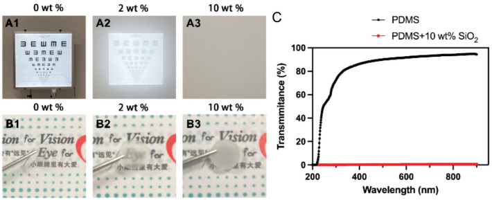
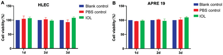
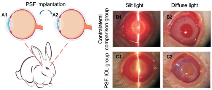
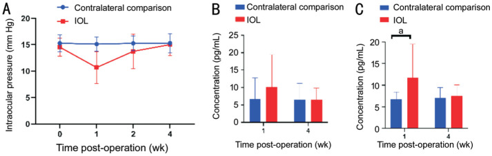
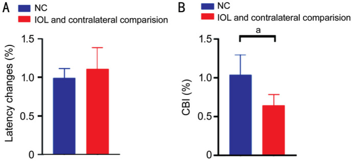
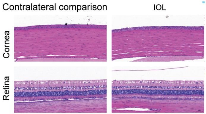
Methods: Poly(dimethyl siloxane)-SiO2 thin films (PSF) with different degrees of opacity as IOL materials were prepared. The light transmission of the PSF-IOL was measured, and its in vitro biosafety was determined by cell counting kit (CCK)-8 assay using the HLEC-B3 cell line and ARPE-19 cell line. Subsequently, the in vivo safety was determined by implanting the PSF-IOL with 10% wt SiO2 into the right eyes of New Zealand white rabbits (PSF-IOL group), and compared with two control groups: contralateral comparison group and normal control (NC) group (Contralateral comparison group: the fellow eye; NC group: a group of binocular normal rabbits without intervention). The flash visual-evoked potentials (F-VEPs) were measured to verify amblyopia.
Results: PSFs containing 0, 2%, and 10% wt SiO2 were successfully constructed. The 0 SiO2 PSF was transparent, while the 10% wt SiO2 PSF was completely opaque. It was found that PSF did not induce unwanted cytotoxicity in HLECs and ARPE19 cells in vitro. In vitro, PSF-IOL with 10% wt SiO2 was also non-toxic, and no significant inflammation or structural changes occurred after four weeks of PSF-IOL implantation. Finally, our IOL-simulated congenital cataract rabbit detected by F-VEPs suggested tentative amblyopia.
Conclusion: A PSF-IOL that mimics cataracts is created. A novel form deprivation model is created by the IOL-simulated congenital cataract rabbit. It can be developed fast and stable and holds great potential for future study.
Keywords: amblyopia; congenital cataract; form deprivation; intraocular lens; monocular deprivation.
International Journal of Ophthalmology Press.
Conflict of interest statement
Conflicts of Interest: Gu SY, None; Xu LM, None; Sun WJ, None; Liang LL, None; Lin L, None; Zou H, None; Xu JY, None; Zheng Y, None; Li YY, None; Zhao YY, None; Chang PJ, None; Zhao YE, None.
Figures
Similar articles
-
Comparison of contact lens and intraocular lens correction of monocular aphakia during infancy: a randomized clinical trial of HOTV optotype acuity at age 4.5 years and clinical findings at age 5 years.JAMA Ophthalmol. 2014 Jun;132(6):676-82. doi: 10.1001/jamaophthalmol.2014.531. JAMA Ophthalmol. 2014. PMID: 24604348 Free PMC article. Clinical Trial.
-
Biosafety of a 3D-printed intraocular lens made of a poly(acrylamide-co-sodium acrylate) hydrogel in vitro and in vivo.Int J Ophthalmol. 2020 Oct 18;13(10):1521-1530. doi: 10.18240/ijo.2020.10.03. eCollection 2020. Int J Ophthalmol. 2020. PMID: 33078100 Free PMC article.
-
Secondary IOL implantation for an aphakic patient with congenital cataract living in Bonin Islands; follow-up study.Strabismus. 2024 Sep;32(3):202-205. doi: 10.1080/09273972.2024.2367068. Epub 2024 Jul 8. Strabismus. 2024. PMID: 38973426
-
Trifocal intraocular lenses versus bifocal intraocular lenses after cataract extraction among participants with presbyopia.Cochrane Database Syst Rev. 2020 Jun 18;6(6):CD012648. doi: 10.1002/14651858.CD012648.pub2. Cochrane Database Syst Rev. 2020. Update in: Cochrane Database Syst Rev. 2023 Jan 27;1:CD012648. doi: 10.1002/14651858.CD012648.pub3. PMID: 32584432 Free PMC article. Updated.
-
[Cataract surgery in children].Med Pregl. 2000 May-Jun;53(5-6):257-61. Med Pregl. 2000. PMID: 11089366 Review. Croatian.
References
LinkOut - more resources
Full Text Sources