

Gastric Carcinomas and Point-of-Care Ultrasound (POCUS): A Report of Two Cases
- PMID: 39697947
- PMCID: PMC11655047
- DOI: 10.7759/cureus.73869
Gastric Carcinomas and Point-of-Care Ultrasound (POCUS): A Report of Two Cases
Abstract
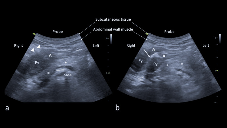
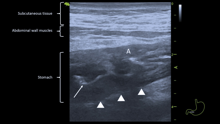
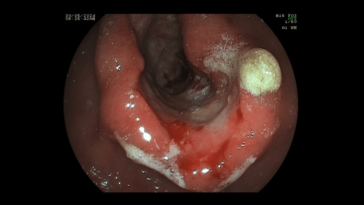
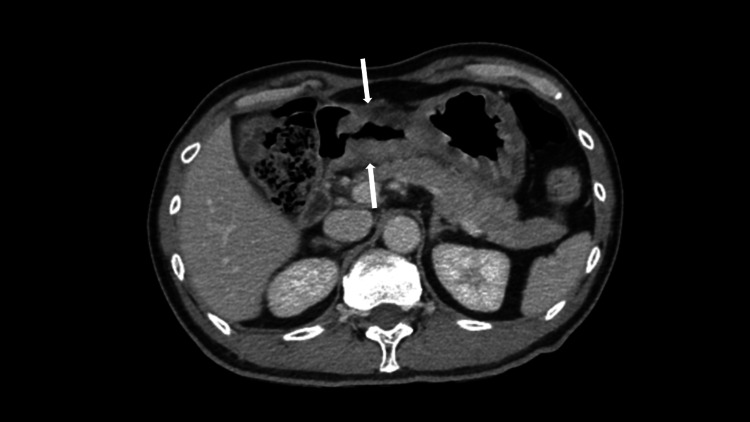
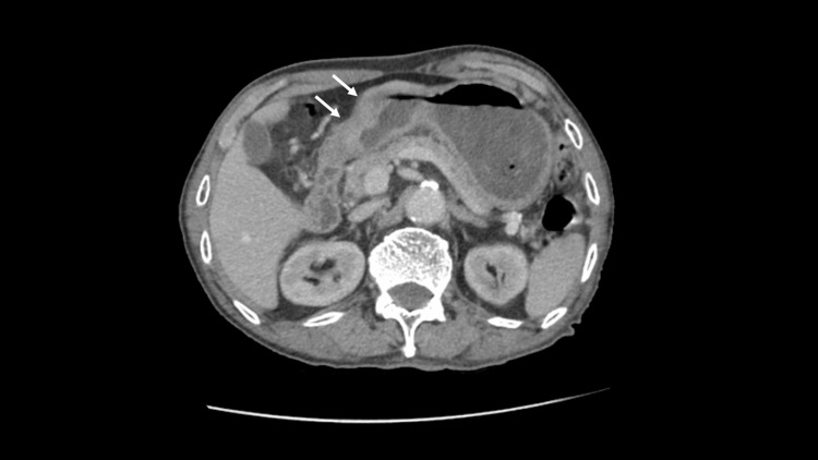
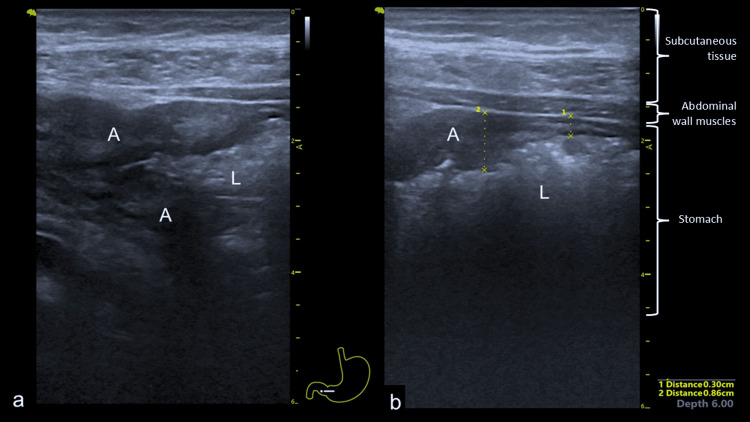
Stomach cancer remains a significant cause of mortality, as most patients are diagnosed at advanced stages. The primary method for diagnosis is endoscopy, along with tissue acquisition, supplemented by endoscopic ultrasound or computed tomography for disease staging. While point-of-care ultrasound (POCUS) is now firmly integrated into clinical practice, it is still not widely utilized. POCUS can be performed at the initial point of contact and provides instant information that can influence investigation strategies. We report two cases of gastric antral carcinoma detected by POCUS, which led to targeted investigations. Both patients underwent expedited upper gastrointestinal endoscopies that confirmed distal gastric carcinoma. These cases highlight the important role of POCUS in triaging patients for timely and appropriate targeted organ investigations.
Keywords: gastric neoplasm; point-of-care-ultrasound; stomach cancer; trans-abdominal ultrasound; ultrasound diagnosis.
Copyright © 2024, Chong et al.
Conflict of interest statement
Human subjects: Consent for treatment and open access publication was obtained or waived by all participants in this study. Conflicts of interest: In compliance with the ICMJE uniform disclosure form, all authors declare the following: Payment/services info: All authors have declared that no financial support was received from any organization for the submitted work. Financial relationships: All authors have declared that they have no financial relationships at present or within the previous three years with any organizations that might have an interest in the submitted work. Other relationships: All authors have declared that there are no other relationships or activities that could appear to have influenced the submitted work.
Figures
References
-
- Gastric cancer. Smyth EC, Nilsson M, Grabsch HI, van Grieken NC, Lordick F. https://www.thelancet.com/journals/lancet/article/PIIS0140-6736(20)31288.... Lancet. 2020;396:635–648. - PubMed
-
- Pre-operative gastric ultrasound in patients at risk of pulmonary aspiration: a prospective observational cohort study. Baettig SJ, Filipovic MG, Hebeisen M, Meierhans R, Ganter MT. Anaesthesia. 2023;78:1327–1337. - PubMed
Publication types
LinkOut - more resources
Full Text Sources