Case Reports
. 2024 Dec 10;29(2):95.
doi: 10.3892/ol.2024.14841. eCollection 2025 Feb.

Metastatic papillary thyroid carcinoma in the soft tissue of the breast in a male patient: A case report

Affiliations
Case Reports

Metastatic papillary thyroid carcinoma in the soft tissue of the breast in a male patient: A case report

Veronika Maráčková et al. Oncol Lett. .

Abstract

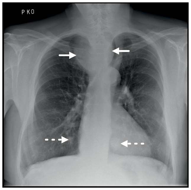
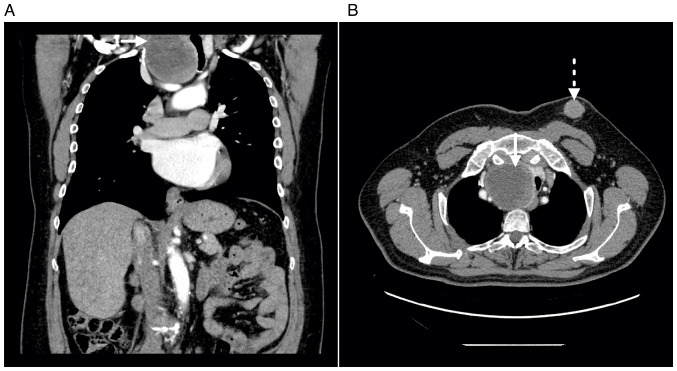
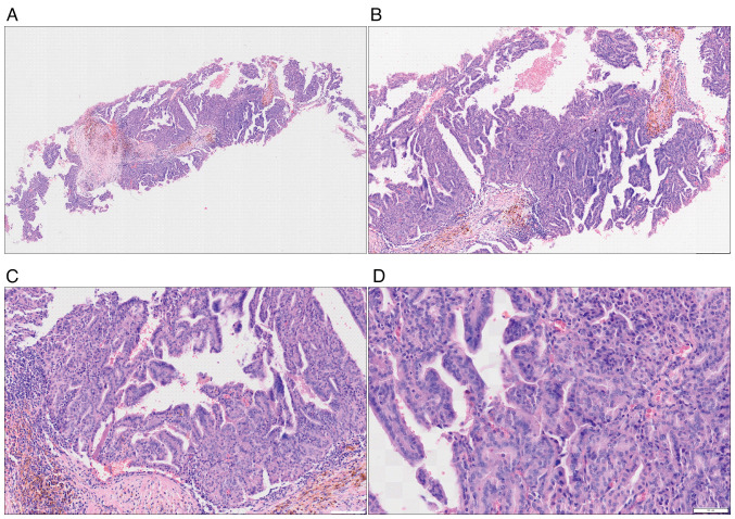
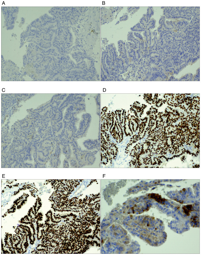
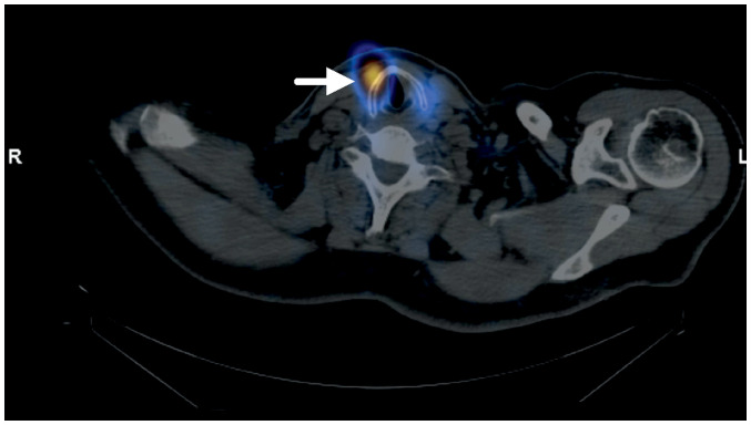
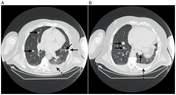
Papillary thyroid carcinoma (PTC) represents ~80% of all thyroid cancers, most frequently presenting in women in the third and fourth decade of life. The first clinical manifestation of PTC commonly includes a palpable mass in the thyroid area or cervical lymphadenopathy in cases of metastatic disease. Hematogenous distant metastases are a sign of an advanced stage of the tumour. The present study reported an extremely rare occurrence of solitary metastasis of a PTC in the left breast of a 63-year-old male patient, mimicking primary male breast cancer (MBC). The presence of a male breast lesion that did not follow the typical imaging criteria for MBC aroused suspicion of a different primary origin. The combination of imaging methods, laboratory findings and fine-needle aspiration techniques enabling cytological and histopathological examination, with the use of a wide panel of immunohistochemical markers, is crucial to establishing a definitive and correct diagnosis.

Keywords: PTC; breast metastasis; mediastinal mass; soft-tissue metastasis.


Conflict of interest statement

The authors declare that they have no competing interests.

Figures

Figure 1.
Figure 1.
Chest X-ray demonstrating a large mass in the upper mediastinum (solid line arrow). The mass has compressed and dislocated the trachea to the left. The biggest lung parenchyma nodulations are observed bilaterally in the lower lungs zones (dashed line arrow).
Figure 2.
Figure 2.
CT results. (A) Coronal plane of enhanced CT scan demonstrating a large hypodense mass with peripheral nodular enhancement on the right side of the upper mediastinum (solid line arrow). The mass is continuous with the right lobe of the thyroid gland, and compresses and causes the trachea to deviate to the left. (B) Axial CT scan demonstrating an irregular lobulated formation in the soft tissue of the left breast with homogenous enhancement following the administration of a contrast medium. The lesion is not in a typical male breast cancer localisation (retroareolar/periareolar) and has smooth margins with fat stranding in the surrounding adipose tissue (dashed line arrow). The large mass on the right side of the upper mediastinum is also highlighted (solid line arrow). CT, computed tomography.
Figure 3.
Figure 3.
Histological findings of the metastasis of papillary thyroid carcinoma, with typical nuclear features and post-haemorrhagic changes in the stroma. Magnification (A) ×20, (B) ×40, (C) ×100 and (D) ×200. All images were captured from the same section of the tissue, which was stained with hematoxylin and eosin.
Figure 4.
Figure 4.
Immunohistochemical markers. Immunohistochemistry results showing absence of (A) gross cystic disease fluid protein 15 (magnification, ×100), (B) GATA binding protein 3 and (C) oestrogen receptor expression in the tumour cells, but positive expression for (D) paired box gene 8, (E) thyroid transcription factor 1 and (F) thyroglobulin (magnification ×100).
Figure 5.
Figure 5.
Whole-body scintigraphy with radioiodine-131 revealing paratracheal tumour residuum (solid line arrow).
Figure 6.
Figure 6.
Unenhanced computed tomography scan in the (A and B) axial planes at different levels demonstrating the increase in the amount and diameter of lung parenchyma nodules bilaterally (solid line arrows). A small amount of pleural effusion is present in the left pleural cavity (dash line arrow).

Similar articles

References

    1. Giuffrida R, Adamo L, Iannolo G, Vicari L, Giuffrida D, Eramo A, Gulisano M, Memeo L, Conticello C. Resistance of papillary thyroid cancer stem cells to chemotherapy. Oncol Lett. 2016;12:687–691. doi: 10.3892/ol.2016.4666. - DOI - PMC - PubMed
    1. Parasuraman L, Kane SV, Pai PS, Shanghvi K. Isolated metastasis in male breast from differentiated thyroid carcinoma - oncological curiosity. A case report and review of literature. Indian J Surg Oncol. 2016;7:91–94. doi: 10.1007/s13193-015-0458-0. - DOI - PMC - PubMed
    1. Shukla N, Osazuwa-Peters N, Megwalu UC. Association between age and nodal metastasis in papillary thyroid carcinoma. Otolaryngol Head Neck Surg. 2021;165:43–49. doi: 10.1177/0194599820966995. - DOI - PubMed
    1. Podolski A, Castelluci E, Halmos B. Precision medicine: BRAF mutations in thyroid cancer. Precis Cancer Med. 2019;2:29. doi: 10.21037/pcm.2019.09.04. - DOI
    1. Lloyd RV, Buehler D, Khanafshar E. Papillary thyroid carcinoma variants. Head Neck Pathol. 2011;5:51–56. doi: 10.1007/s12105-010-0236-9. - DOI - PMC - PubMed

Publication types

LinkOut - more resources