Review
. 2023 May 23;4(2):218-239.
doi: 10.20517/evcna.2023.19. eCollection 2023.

Bioprinting extracellular vesicles as a "cell-free" regenerative medicine approach

Affiliations
Review

Bioprinting extracellular vesicles as a "cell-free" regenerative medicine approach

Kexin Jiao et al. Extracell Vesicles Circ Nucl Acids. .

Abstract

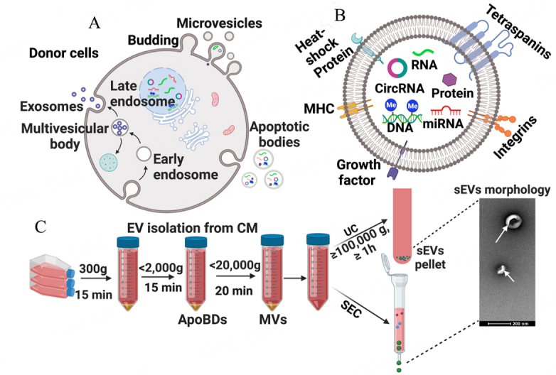
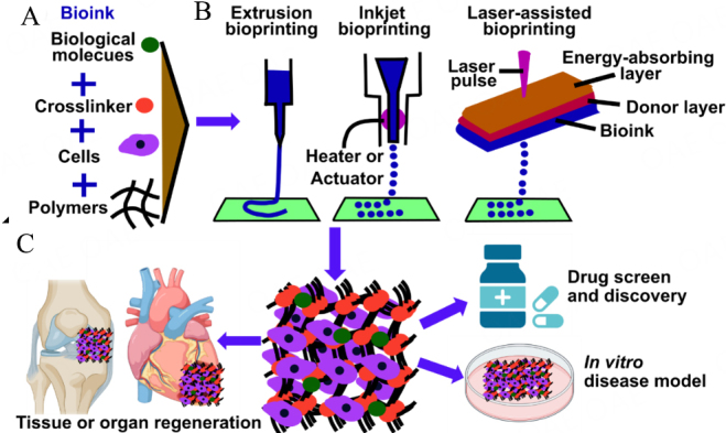
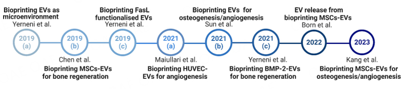
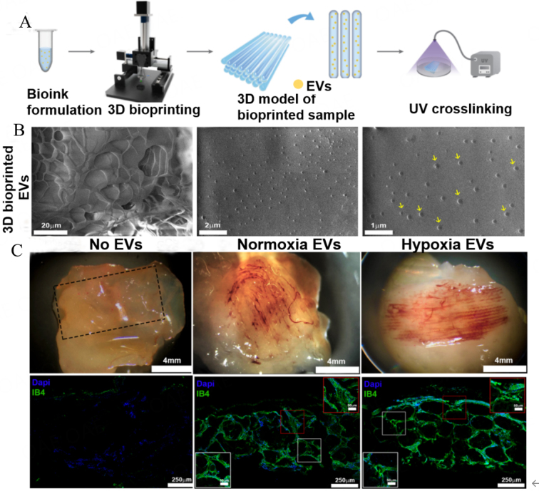
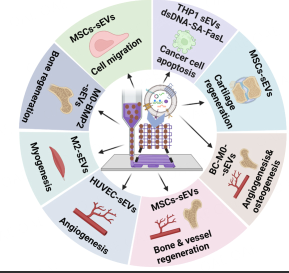
Regenerative medicine involves the restoration of tissue or organ function via the regeneration of these structures. As promising regenerative medicine approaches, either extracellular vesicles (EVs) or bioprinting are emerging stars to regenerate various tissues and organs (i.e., bone and cardiac tissues). Emerging as highly attractive cell-free, off-the-shelf nanotherapeutic agents for tissue regeneration, EVs are bilayered lipid membrane particles that are secreted by all living cells and play a critical role as cell-to-cell communicators through an exchange of EV cargos of protein, genetic materials, and other biological components. 3D bioprinting, combining 3D printing and biology, is a state-of-the-art additive manufacturing technology that uses computer-aided processes to enable simultaneous patterning of 3D cells and tissue constructs in bioinks. Although developing an effective system for targeted EVs delivery remains challenging, 3D bioprinting may offer a promising means to improve EVs delivery efficiency with controlled loading and release. The potential application of 3D bioprinted EVs to regenerate tissues has attracted attention over the past few years. As such, it is timely to explore the potential and associated challenges of utilizing 3D bioprinted EVs as a novel "cell-free" alternative regenerative medicine approach. In this review, we describe the biogenesis and composition of EVs, and the challenge of isolating and characterizing small EVs - sEVs (< 200 nm). Common 3D bioprinting techniques are outlined and the issue of bioink printability is explored. After applying the following search strategy in PubMed: "bioprinted exosomes" or "3D bioprinted extracellular vesicles", eight studies utilizing bioprinted EVs were found that have been included in this scoping review. Current studies utilizing bioprinted sEVs for various in vitro and in vivo tissue regeneration applications, including angiogenesis, osteogenesis, immunomodulation, chondrogenesis and myogenesis, are discussed. Finally, we explore the current challenges and provide an outlook on possible refinements for bioprinted sEVs applications.

Keywords: 3D bioprinting; bioprinted sEVs; regenerative medicine; small extracellular vesicles.


Conflict of interest statement

All authors declared that there are no conflicts of interest.

Figures

Figure 1
Figure 1
EVs Biogenesis, components, and cell-derived EVs solation method. (A) Biogenesis of Extracellular vehicles (EVs); (B) components of EVs; (C) Common EVs isolation steps using a serial centrifuge and sEVs isolation by either UC or SEC method prior to TEM analysis of sEVs morphology. MHC: Major histocompatibility complex; CM: condition media; ApoBDs: Apoptotic bodies; MVs: Multivesicular body; SEC: Size Exclusion Chromatography; UC: ultracentrifugation.
Figure 2
Figure 2
Schematics of bioinks (A), 3D bioprinting methods (B) and the application of 3D bioprinting (C). (A) Bioink typically includes biological molecules, live cells, cross-linker, and polymers (GelMA hydrogel). (B) Current widely used 3D bioprinting techniques of extrusion, inkjet and laser-assisted bioprinting. (C) Three main applications of 3D bioprinting lie in drug screening, in vitro disease models and tissue regeneration.
Figure 3
Figure 3
This review includes 8 research studies, as demonstrated by the timeline.
Figure 4
Figure 4
Bioprinted HUVECs-sEVs constructs promote angiogenesis (modified from [103]). (A) Schematic representation of the bioprinting process to manufacture EV-loaded scaffolds; (B) SEM images of 3D bioprinted GelMA with HUVECs-sEVs. Yellow arrows denote the EVs; (C) Bioprinted HUVECs-sEVs promote in vivo vessel formation after subcutaneous implantation in immunocompromised mice, with IB4-positive blood vessel structures. IB4, Isolectin B4; Normoxia EVs, EVs from normoxia HUVECs; hypoxia EVs, EVs from hypoxia HUVECs.
Figure 5
Figure 5
In vitro and in vivo assessment of bioprinted BMP2-EVs (modified from[105]). (A) Bioprinted patterns of Alexa Fluor 488-labeled BMP2 (green) loaded in PKH26-labeled EVs (red); (B) ALP staining of C2C12 cells at 72 h post-seeding on bioprinted BMP2-EVs patterns with indicated OPs; (C) Representative microCT 3D reconstructions of mouse leg scans containing either native EVs or BMP2-EVs bioprinted implants. Arrow points to heterotopic ossification; (D) Representative histological images showing H&E and Masson's trichrome staining of native EVs and BMP2-EVs bioprinted implants (*indicates bone tissue).
Figure 6
Figure 6
Bioprinted MSCs-sEVs constructs facilitate both cartilage and bone regeneration in a rabbit (modified from [108]). (A) Schematic illustration of stereolithography-based bioprinted MSCs-sEVs in decellularized cartilage ECM and GelMA bioink prior to in vivo osteochondral defect implantation in a rabbit. SEV is known as sEVs; (B) Bioprinted MSCs-sEVs promoted both cartilage and bone formation after 6 and 12 weeks of implantation. Group I, osteochondral defect only; Group II, 3D bioprinted GelMA; Group III, 3D bioprinted ECM/GelMA scaffold; Group IV, 3D printed ECM/GelMA/sEVs scaffold.
Figure 7
Figure 7
Summary of sEVs from different cell sources and functions of bioprinted sEVs as "cell-free" regenerative medicine approaches. MSC-sEVs: MSC-derived small EVs; HUVEC-sEVs: Human umbilical vein endothelial cell-derived small EVs; M0-BMP2-sEVs: inactivated macrophage (M0) derived sEVs loaded with BMP2 protein; M2-sEVs: Macrophage stage 2 small EV (pro-regenerative); THP1-sEVs-dsDNA-SA-FasL: human monocyte cell line THP1 derived sEVs tettered with dsDNA and modified with streptavidin (SA) and Fas Ligand (FasL); BC-M0-sEVs: bioceramic-induced macrophage-derived sEVs.

Similar articles

Cited by

References

    1. Schäfer N, Grässel S. New refinements aim to optimize articular cartilage tissue engineering. Nat Rev Rheumatol. 2023;19:66–7. doi: 10.1038/s41584-022-00889-y. - DOI - PubMed
    1. Wang X, Chen J, Tian W. Strategies of cell and cell-free therapies for periodontal regeneration: the state of the art. Stem Cell Res Ther. 2022;13:536. doi: 10.1186/s13287-022-03225-z. - DOI - PMC - PubMed
    1. Safdar A, Saleem A, Tarnopolsky MA. The potential of endurance exercise-derived exosomes to treat metabolic diseases. Nat Rev Endocrinol. 2016;12:504–17. doi: 10.1038/nrendo.2016.76. - DOI - PubMed
    1. Théry C, Witwer KW, Aikawa E, et al. Minimal information for studies of extracellular vesicles 2018 (MISEV2018): a position statement of the International Society for Extracellular Vesicles and update of the MISEV2014 guidelines. J Extracell Vesicles. 2018;7:1535750. doi: 10.1080/20013078.2018.1535750. - DOI - PMC - PubMed
    1. Niel G, D'Angelo G, Raposo G. Shedding light on the cell biology of extracellular vesicles. Nat Rev Mol Cell Biol. 2018;19:213–28. doi: 10.1038/nrm.2017.125. - DOI - PubMed

LinkOut - more resources