. 2024 Nov 28:8:100159.
doi: 10.1016/j.crstbi.2024.100159. eCollection 2024.

Pathogenic single nucleotide polymorphisms in RhoA gene: Insights into structural and functional impacts on RhoA-PLD1 interaction through molecular dynamics simulation

Affiliations

Pathogenic single nucleotide polymorphisms in RhoA gene: Insights into structural and functional impacts on RhoA-PLD1 interaction through molecular dynamics simulation

Mahbub Hasan et al. Curr Res Struct Biol. .

Abstract

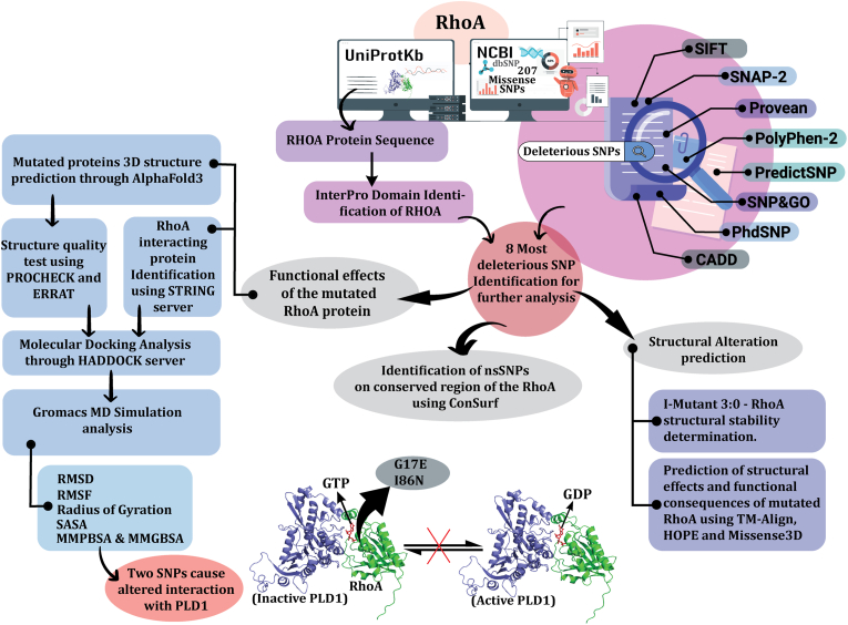
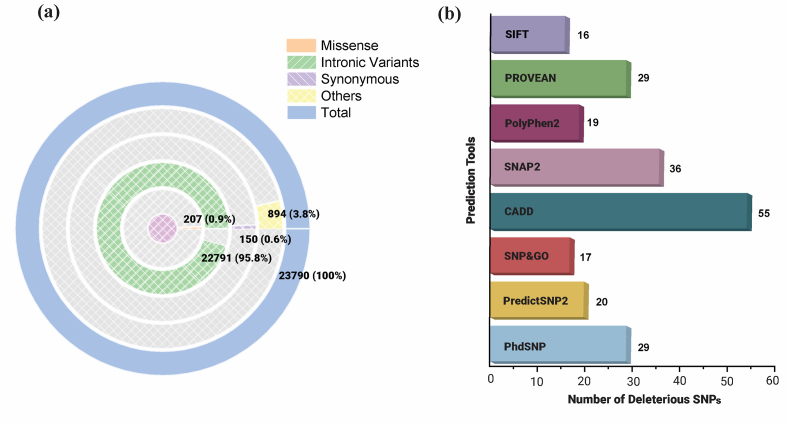
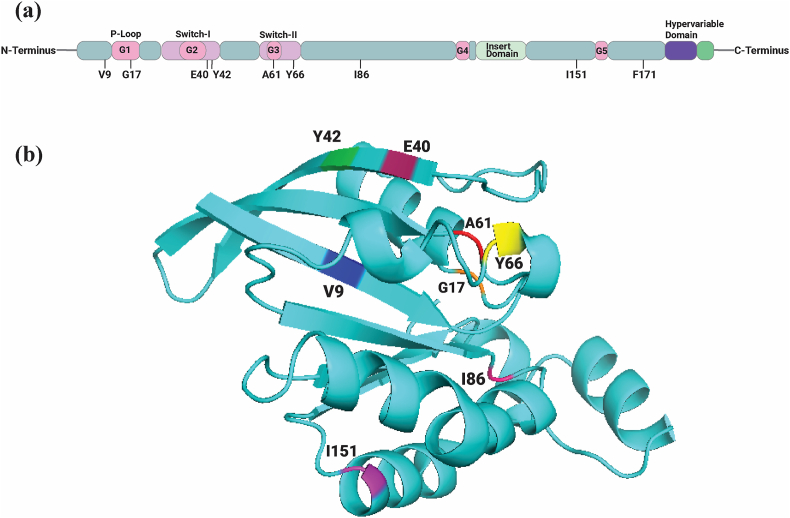
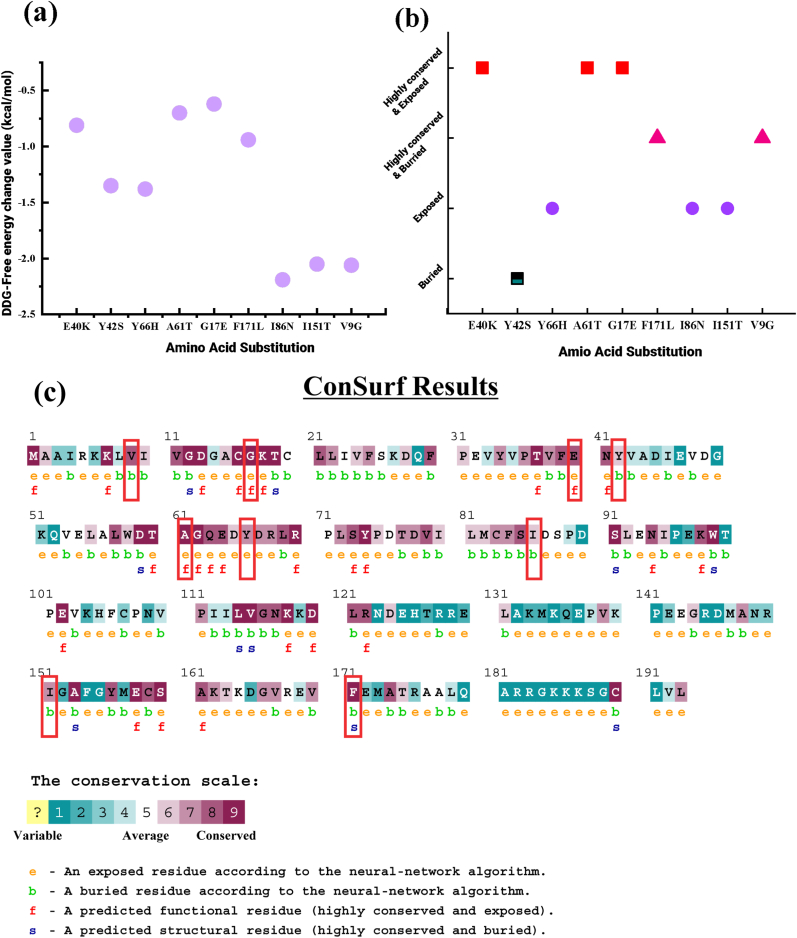
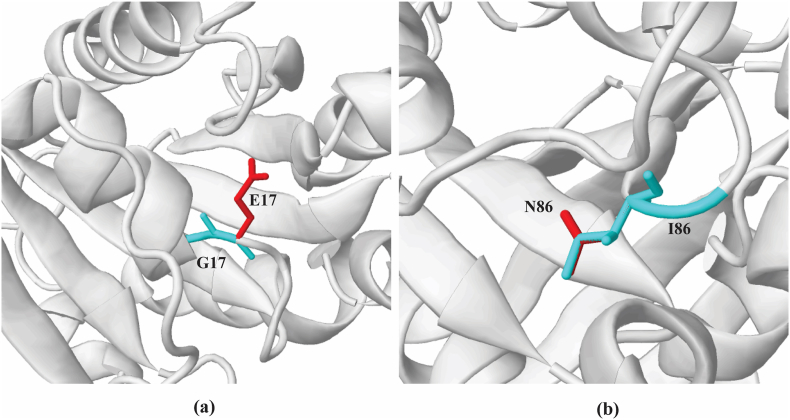
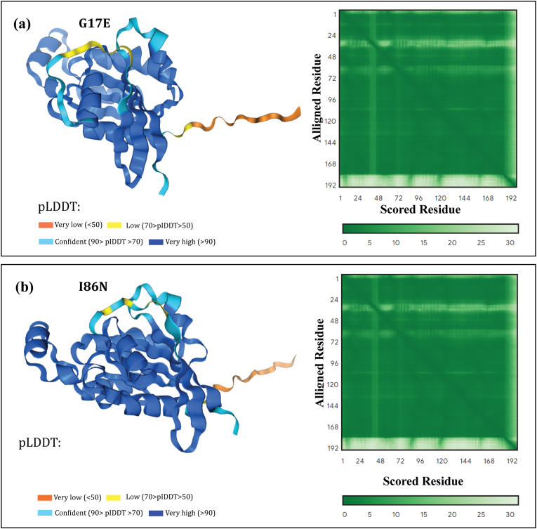
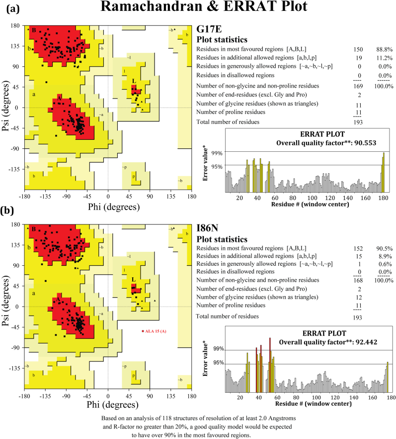
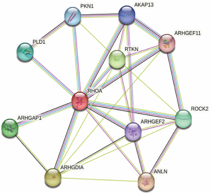
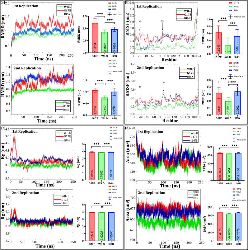
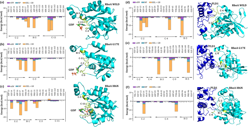
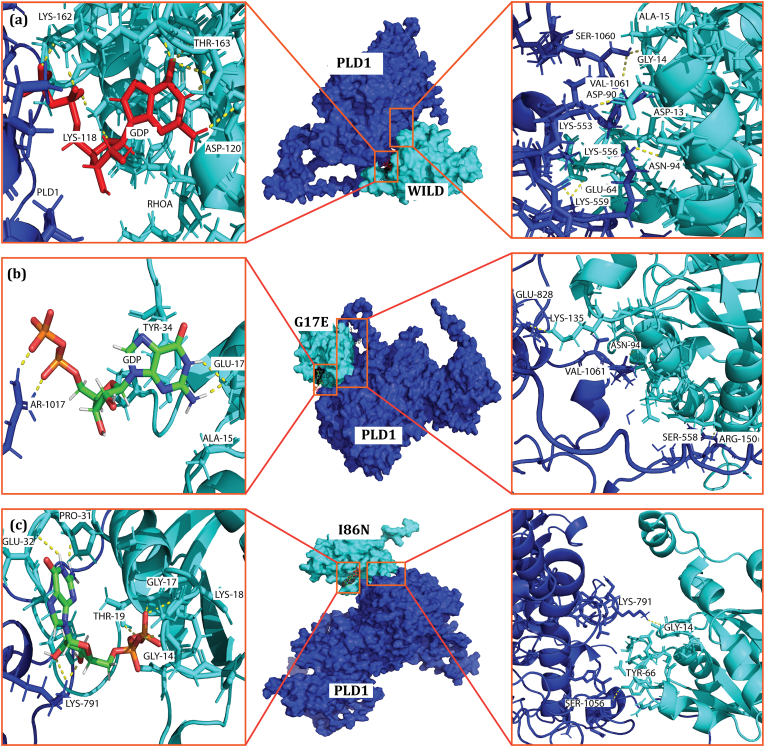
Molecular switches serve as key regulators of biological systems by acting as one of the crucial driving forces in the initiation of signal transduction pathway cascades. The Ras homolog gene family member A (RhoA) is one of the molecular switches that binds with GTP in order to cycle between an active GTP-bound state and an inactive GDP-bound state. Any aberrance in control over this circuit, particularly due to any perturbation in switching, leads to the development of different pathogenicity. Consequently, the single nucleotide polymorphisms (SNPs) within the RhoA gene, especially deleterious genetic variations, are crucial to study to forecast structural alteration and their functional impacts in light of disease onset. In this comprehensive study, we employed a range of computational tools to screen the deleterious SNPs of RhoA from 207 nonsynonymous SNPs (nsSNPs). By utilizing 7 distinct tools for further analysis, 8 common deleterious SNPs were sorted, among them 5 nsSNPs (V9G, G17E, E40K, A61T, F171L) were found to be in the highly conserved regions, with E40K and A61T at G2 and G3 motif of the GTP-binding domain respectively, indicating potential perturbation in GTP/GDP binding ability of the protein. RhoA-GDP complex interacts with the enzyme phospholipase, specifically PLD1, to regulate different cellular activities. PLD1 is also a crucial regulator of thrombosis and cancer. In that line of focus, our initial structural analysis of Y66H, A61T, G17E, I86N, and I151T mutations of RhoA revealed remarkable decreased hydrophobicity from which we further filtered out G17E and I86N which may have potential impact on the RhoA-GDP-PLD1 complex. Intriguingly, the comparative 250 ns (ns) molecular dynamics (MD) simulation of these two mutated complexes revealed overall structural instability and altered interaction patterns. Therefore, further investigation into these deleterious mutations with in vitro and in vivo studies could lead to the identification of potential biomarkers in terms of different pathogenesis and could also be utilized in personalized therapeutic targets in the long run.

Keywords: Deleterious SNP; Molecular dynamic simulation; Molecular switching; RhoA; RhoA-GDP-PLD1.


Conflict of interest statement

The authors declare that they have no known competing financial interests or personal relationships that could have appeared to influence the work reported in this paper.

Figures

Fig. 1
Fig. 1
Schematic representation of the most deleterious SNP identification of RhoA gene and their subsequent effect analysis.
Fig. 2
Fig. 2
In silico-based identification of deleterious SNPs in the RhoA gene. (a) A circular diagram highlighting the various types of SNPs in RhoA. (b) The total number of missense SNPs and predicted deleterious SNPs by various algorithms are described in the bar plot.
Fig. 3
Fig. 3
In the human RhoA crystal structure, the structural domains and motifs present in RhoA are highlighted in a representative three-dimensional structure (a), and the identified variant position for 8 nsSNPs is labeled in a cartoon representation (b).
Fig. 4
Fig. 4
Effect of structural stability and evolutionary conservation analysis of protein residues (a) The effects of nsSNPs on protein stability predicted by I-MUTANT 3.0 based on structural stability in terms of RI and free energy change value (DDG) are plotted. ConSurf results are highlighted in red (b) and revealed that 5 out of 9 nsSNPs are highly conserved which is plotted in (c).
Fig. 5
Fig. 5
Structural alteration of the wild-type residue by the mutant G17E (a) and I86N (b) illustrated by Missense 3D. The wild-type residue is presented as cyan, and the mutant residue is shown in red.
Fig. 6
Fig. 6
3D protein modeling with AlphaFold3. AlphaFold3 predicted mutated structure of RhoA (a) G17E, and (b) I86N. The lower pLDDT score (the orange and yellow region) indicates disordered region. Both models here have very high and confidence scores across lion's share of the structures, making the structures highly reliable. The lower the PAE value, the better defined the residue's relative position is assumed to be, however when the color changes to light green from dark green, the PAE score rises, and their relative positions are anticipated with less confidence. The predicted template modeling (pTM) score for G17E is 0.89 and for I86N is 0.9. A score above 0.5 predicts that the folds of the model is possibly similar to the true structure.
Fig. 7
Fig. 7
Predicted structure validation. Ramachandran and ERRAT plots were generated using SAVES server for the mutated RhoA proteins G17E and I86N. The number of residues in the most favored regions are 150 (88.8%) for G17E and 152 (90.5%) for I86N, with no residues found in the disallowed regions in both cases. The overall quality factor for the RhoA G17E and I86N in the ERRAT plot is 90.553 and 92.442, respectively.
Fig. 8
Fig. 8
The STRING web server generates proteins that interact with RhoA proteins in various biological pathways.
Fig. 9
Fig. 9
Molecular dynamics analysis of wild-type and mutant (G17E and I86N) RhoA proteins. The Root Mean Square Deviation (RMSD), Root Mean Square Fluctuation (RMSF), Radius of Gyration (Rg), and Solvent Accessible Surface Area (SASA) plots are shown for two replicates. (Top left) RMSD plots reveal stability differences among wild-type, G17E, and I86N mutants over 250 ns, with G17E showing higher deviation, indicating reduced stability. (Top right) RMSF plots highlight residue flexibility, with G17E and I86N showing more fluctuation, particularly around residues 60–90 and 180–193. (Bottom left) Rg plots display the compactness of the proteins, with G17E being more expanded. (Bottom right) SASA plots indicate solvent exposure, with the G17E mutant showing the largest surface area, further suggesting a looser structure. Statistical significance is indicated with asterisks.
Fig. 10
Fig. 10
Interaction free energy analysis of (a, b, c) Wild-type RhoA, G17E, and I86N mutant residues interacting with GDP, and (d, e, f) Wild-type RhoA, G17E, and I86N mutant residues interacting with PLD1 within a 4 Å binding vicinity. The left panels show the comparison of interaction free energy components (vdW, NP, EEL + GB) for RhoA-GDP complexes, and the right panels display the same for RhoA-PLD1 complexes after 250 ns of molecular dynamics simulations. The most significant residues contributing to the binding free energy in both complexes are highlighted in yellow within the structural representations.
Fig. 11
Fig. 11
Structural representation of interactions between wild-type and mutant (G17E and I86N) RhoA proteins with GDP and PLD1. Panels (a), (b), and (c) show the detailed interactions of key residues within the binding sites of wild-type, G17E, and I86N RhoA with GDP (left) and PLD1 (right), respectively. Significant residues involved in the interactions are labeled, and hydrogen bonds are shown as yellow dashed lines. The surface representation highlights the interaction interface between RhoA (cyan) and PLD1 (blue), with zoomed-in views emphasizing critical residues contributing to the binding between these proteins.

Similar articles

References

    1. Abid F., Khan K., Badshah Y., Ashraf N.M., Shabbir M., Hamid A., Afsar T., Almajwal A., Razak S. Non-synonymous SNPs variants of PRKCG and its association with oncogenes predispose to hepatocellular carcinoma. Cancer Cell Int. 2023;23:1–24. doi: 10.1186/S12935-023-02965-Z. - DOI - PMC - PubMed
    1. Abraham M.J., Murtola T., Schulz R., Páll S., Smith J.C., Hess B., Lindah E. GROMACS: high performance molecular simulations through multi-level parallelism from laptops to supercomputers. SoftwareX. 2015;1–2:19–25. doi: 10.1016/J.SOFTX.2015.06.001. - DOI
    1. Abramson J., Adler J., Dunger J., Evans R., Green T., Pritzel A., Ronneberger O., Willmore L., Ballard A.J., Bambrick J., Bodenstein S.W., Evans D.A., Hung C.C., O'Neill M., Reiman D., Tunyasuvunakool K., Wu Z., Žemgulytė A., Arvaniti E., Beattie C., Bertolli O., Bridgland A., Cherepanov A., Congreve M., Cowen-Rivers A.I., Cowie A., Figurnov M., Fuchs F.B., Gladman H., Jain R., Khan Y.A., Low C.M.R., Perlin K., Potapenko A., Savy P., Singh S., Stecula A., Thillaisundaram A., Tong C., Yakneen S., Zhong E.D., Zielinski M., Žídek A., Bapst V., Kohli P., Jaderberg M., Hassabis D., Jumper J.M. Accurate structure prediction of biomolecular interactions with AlphaFold 3. Nature. 2024;630:493–500. doi: 10.1038/s41586-024-07487-w. 2024 630:8016. - DOI - PMC - PubMed
    1. Adzhubei I.A., Schmidt S., Peshkin L., Ramensky V.E., Gerasimova A., Bork P., Kondrashov A.S., Sunyaev S.R. A method and server for predicting damaging missense mutations. Nat. Methods. 2010;7:248–249. doi: 10.1038/nmeth0410-248. 2010 7:4. - DOI - PMC - PubMed
    1. Al-nakhle H.H., Khateb A.M. Comprehensive in silico characterization of the coding and non-coding SNPs in human dectin-1 gene with the potential of high-risk pathogenicity associated with fungal infections. Diagnostics. 2023;13:1785. doi: 10.3390/DIAGNOSTICS13101785/S1. - DOI - PMC - PubMed

LinkOut - more resources