

Alterations in large-scale resting-state network nodes following transcranial focused ultrasound of deep brain structures
- PMID: 39698148
- PMCID: PMC11652661
- DOI: 10.3389/fnhum.2024.1486770
Alterations in large-scale resting-state network nodes following transcranial focused ultrasound of deep brain structures
Abstract
Background: Low-intensity transcranial focused ultrasound (tFUS) is a brain stimulation approach that holds promise for the treatment of brain-based disorders. Studies in humans have shown that tFUS can successfully modulate perfusion in focal sonication targets, including the amygdala; however, limited research has explored how tFUS impacts large-scale neural networks.
Objective: The aim of the current study was to address this gap and examine changes in resting-state connectivity between large-scale network nodes using a randomized, double-blind, within-subjects crossover study design.
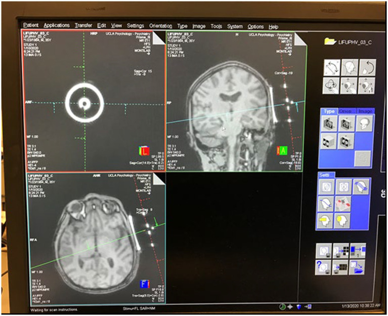
Methods: Healthy adults (n = 18) completed two tFUS sessions, 14 days apart. Each session included tFUS of either the right amygdala or the left entorhinal cortex (ErC). The inclusion of two active targets allowed for within-subjects comparisons as a function of the locus of sonication. Resting-state functional magnetic resonance imaging was collected before and after each tFUS session.
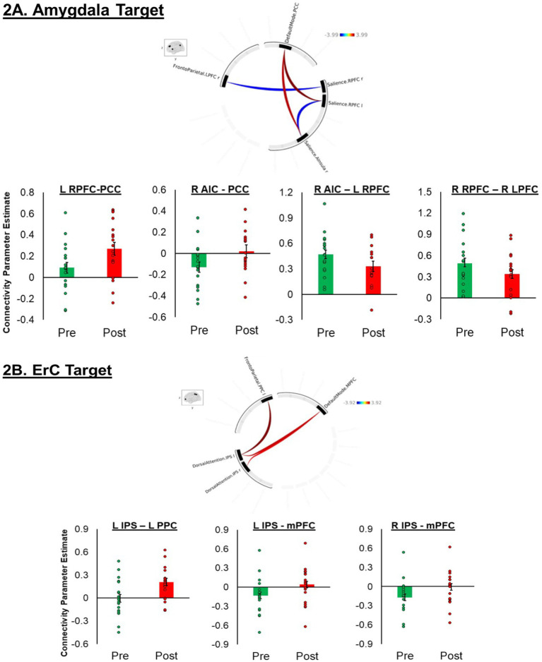
Results: tFUS altered resting-state functional connectivity (rsFC) within and between rs-network nodes. Pre-to-post sonication of the right amygdala modulated connectivity within nodes of the salience network (SAN) and between nodes of the SAN and the default mode network (DMN) and frontoparietal network (FRP). A decrease in SAN to FPN connectivity was specific to the amygdala target. Pre-to-post sonication of the left ErC modulated connectivity between the dorsal attention network (DAN) and FPN and DMN. An increase in DAN to DMN connectivity was specific to the ErC target.
Conclusion: These preliminary findings may suggest that tFUS induces neuroplastic changes beyond the immediate sonication target. Additional studies are needed to determine the long-term stability of these effects.
Keywords: amygdala; default mode network; entorhinal cortex; resting-state functional connectivity; salience network (SN); transcranial focused ultrasound.
Copyright © 2024 Gorka, Jimmy, Koning, Phan, Rotstein, Hoang-Dang, Halavi, Spivak, Monti, Reggente, Bookheimer and Kuhn.
Conflict of interest statement
The authors declare that the research was conducted in the absence of any commercial or financial relationships that could be construed as a potential conflict of interest.
Figures
References
LinkOut - more resources
Full Text Sources
Miscellaneous