Review
. 2023 Mar 9;4(1):27-43.
doi: 10.20517/evcna.2023.05. eCollection 2023.

Hijacking intercellular trafficking for the spread of protein aggregates in neurodegenerative diseases: a focus on tunneling nanotubes (TNTs)

Affiliations
Review

Hijacking intercellular trafficking for the spread of protein aggregates in neurodegenerative diseases: a focus on tunneling nanotubes (TNTs)

Ranabir Chakraborty et al. Extracell Vesicles Circ Nucl Acids. .

Abstract

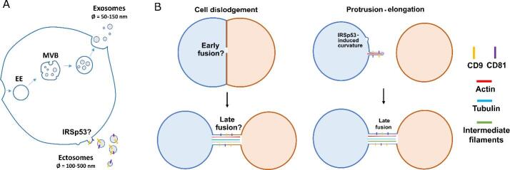
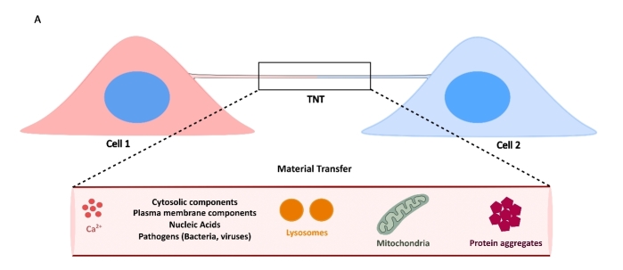
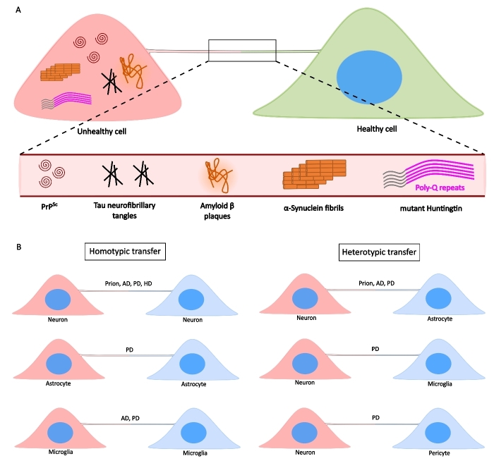
Over the years, the influence of secretory mechanisms on intercellular communication has been extensively studied. In the central nervous system (CNS), both trans-synaptic (neurotransmitter-based) and long-distance (extracellular vesicles-based) communications regulate activities and homeostasis. In less than a couple of decades, however, there has been a major paradigm shift in our understanding of intercellular communication. Increasing evidence suggests that besides secretory mechanisms (via extracellular vesicles), several cells are capable of establishing long-distance communication routes referred to as Tunneling Nanotubes (TNTs). TNTs are membranous bridges classically supported by F-Actin filaments, allowing for the exchange of different types of intracellular components between the connected cells, ranging from ions and organelles to pathogens and toxic protein aggregates. The roles of TNTs in pathological spreading of several neurodegenerative conditions such as Prion diseases, Alzheimer's disease (AD), Parkinson's disease (PD), and Huntington's disease (HD) have been well established. However, the fragile nature of these structures and lack of specific biomarkers raised some skepticism regarding their existence. In this review, we will first place TNTs within the spectrum of intercellular communication mechanisms before discussing their known and hypothesized biological relevance in vitro and in vivo in physiological and neurodegenerative contexts. Finally, we discuss the challenges and promising prospects in the field of TNT studies.

Keywords: Tunneling nanotubes; intercellular communication; neurodegenerative diseases.


Conflict of interest statement

All authors declared that there are no conflicts of interest.

Figures

Figure 1
Figure 1
Formation mechanisms of EVs and TNTs. (A) EVs are generated via different pathways. Generation of exosomes require inner budding within EE maturing into MVBs, whereas ectosomes form via negative membrane curvature-induced budding at the plasma membrane. Both the tetraspanins CD9 and CD81 are components of ectosomal membranes and are found on TNTs. (B) TNTs can be formed via different mechanisms, viz. cell dislodgement (left panels), wherein cells that come in contact with each other leave behind a tubular connection when they move apart, and protrusion-elongation (right panels), where one cell, following negative membrane curvature, actively extends a protrusion towards a neighbouring cell to eventually form a functional connection.
Figure 2
Figure 2
Functional nature of TNTs is defined by their ability to facilitate material transfer between connected cells. A: Cells connected by TNTs can allow for exchange of various intracellular materials, such as ions, cytosolic and plasma membrane components, nucleic acids such as messenger and/or regulatory RNAs, organelles such as lysosomes and mitochondria, and cytotoxic protein aggregates.
Figure 3
Figure 3
TNTs facilitate the transfer of NDs-causing protein aggregates. A: Unhealthy cells containing protein aggregates can form TNTs with a naïve, healthy cell, eventually spreading aggregates such as PrPSc (Prion’s disease), Tau and Amyloid-β (AD), α-Syn fibrils (PD), and mHtt (HD). B: Such transfers can happen between the same type of cells via homotypic TNTs (left panels), or between different cell types via heterotypic TNTs (right panels).

Similar articles

Cited by

References

    1. Handly LN, Pilko A, Wollman R. Paracrine communication maximizes cellular response fidelity in wound signaling. Elife. 2015;4:e09652. doi: 10.7554/eLife.09652. - DOI - PMC - PubMed
    1. Sánchez G, Bunn KE, Pua HH, Rafat M. Extracellular vesicles: mediators of intercellular communication in tissue injury and disease. Cell Commun Signal. 2021;19:104. doi: 10.1186/s12964-021-00787-y. - DOI - PMC - PubMed
    1. Cocucci E, Meldolesi J. Ectosomes and exosomes: shedding the confusion between extracellular vesicles. Trends Cell Biol. 2015;25:364–72. doi: 10.1016/j.tcb.2015.01.004. - DOI - PubMed
    1. Gould SJ, Booth AM, Hildreth JE. The Trojan exosome hypothesis. Proc Natl Acad Sci USA. 2003;100:10592–7. doi: 10.1073/pnas.1831413100. - DOI - PMC - PubMed
    1. Vella LJ, Sharples RA, Lawson VA, Masters CL, Cappai R, Hill AF. Packaging of prions into exosomes is associated with a novel pathway of PrP processing. J Pathol. 2007;211:582–90. doi: 10.1002/path.2145. - DOI - PubMed

LinkOut - more resources