Review
. 2023 Mar 15;4(1):44-58.
doi: 10.20517/evcna.2023.03. eCollection 2023.

Four distinct cytoplasmic structures generate and release specific vesicles, thus opening the way to intercellular communication

Affiliations
Review

Four distinct cytoplasmic structures generate and release specific vesicles, thus opening the way to intercellular communication

Gabriella Racchetti et al. Extracell Vesicles Circ Nucl Acids. .

Abstract

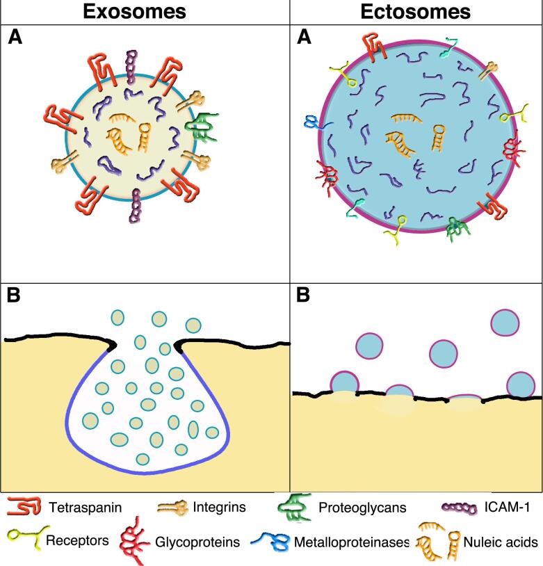
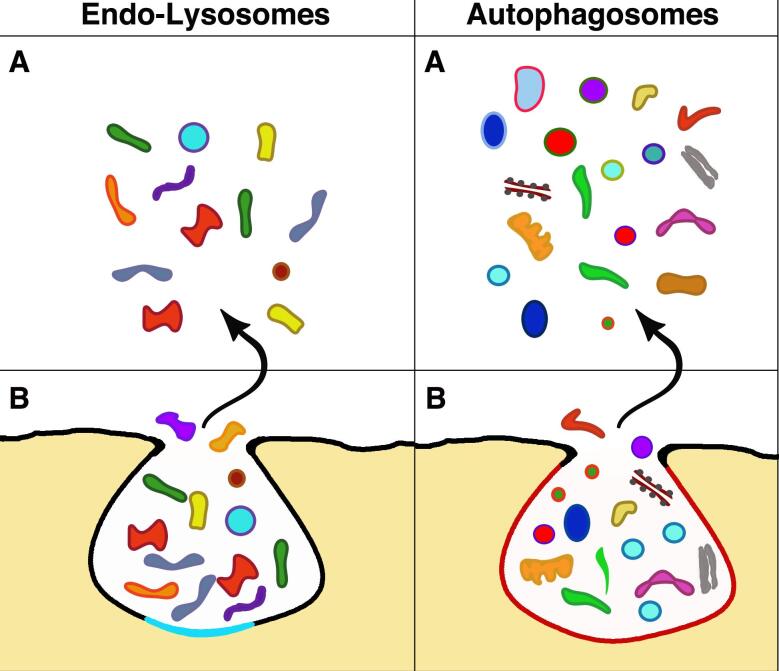
In all cells, generation and release of specific vesicles are the initial steps of back-and-forth intercellular communication. These processes are critical in normal physiology and pathophysiology. Vesicles have particular functions appropriate to their targets. When stimulated, they are released into the extracellular space. Four cytoplasmic membrane-bound structures generate their particular vesicles. Among these structures, multivesicular bodies (MVBs) can accumulate many small vesicles in their lumen; release occurs upon MVB exocytosis. Ectosomes are larger vesicles characterized by their responses and are generated directly and released independently from specific microdomains pre-established in the thickness of the plasma membrane. Most lysosomes do not generate vesicles. However, unique components of a minor form, the endo-lysosome, constitute the third class of structures that release a few vesicles by exocytosis with molecules and structures inducing changes in the extracellular environment. The autophagosome, the fourth structure, releases several heterogeneous vesicles by exocytosis with malformed bio-molecules, assembled structures, and damaged organelles. Interestingly, the frequent interaction of autophagosomes with MVBs and their exosomes contributes to the regulation and intensity of their action. The specificity and function of released vesicles depend on their membranes' and luminal cargoes' composition and dynamics. An ongoing investigation of the various vesicles reveals new properties regarding their generation, release, and resulting extracellular processes. The growth of information about structures and their vesicles progressively extends the knowledge base regarding cell communication and contributes to their clinical applications.

Keywords: Autophagosomes; cargo; ectosomes and exosomes; endo-lysosomes; endocytosis; exocytosis; extracellular vesicles (EVs); lysosome storage disorders (LSDs); membrane fusions; microdomains; multivesicular bodies (MVBs); unconventional protein secretion (UPS).


Conflict of interest statement

Authors declared that there are no conflicts of interest.

Figures

Figure 1
Figure 1
Structure and composition of the two vesicles; their release from the plasma membrane by exocytosis and outward budding. A comparison of the images confirms that exosome vesicles are smaller than ectosome vesicles. Plasma membranes of exosomes are in black, whereas those of vesicle membranes are different: sky blue for those of endocytic nature, i.e., the MVB (B) and its exosomes (A and B); violet for those of ectosomes (A and B), in which plasma membrane and endosome are mixed. In exosome and ectosome vesicles, the color of the lumen is substantially different: lemon yellow for exosomes (A and B) and maya blue for ectosomes (A and B). The present comparison emphasizes the moderate chemical distinction between the vesicle types (A). Several essential membrane proteins are listed below. Some (e.g., tetraspanins and integrins) predominate in exosome but are also present in ectosome. In contrast, other proteins (e.g., receptors, glycoproteins, and metalloproteinases) are present in ectosomes and are not appreciable in the exosome. In both A vesicles, nucleic acids are present (neon orange) in the depth of the lumen. B images show the release of the two vesicle types: by exocytosis of MVBs, with the subsequent release of exosomes, by outward budding of ectosomes followed by growth and then outward release from the plasma membrane. The images of the two enlarged vesicles shown in two A are reproduced with permission[26].
Figure 2
Figure 2
Exocytosis of the two organelles (B) and their released material spread in the extracellular space (A). Analogous to the exosomes in Figure 1, these images of an endo-lysosome and an autophagosome illustrate exocytoses with release to the extracellular space. However, the two exocytosis forms differ concerning exosomes in Figure 1 and from one another. The endo-lysosome B image is delimited by a membrane combination induced by the fusion of a lysosome (black, the same color as the plasma membrane) with an endocytic cisterna (sky blue). The other B comes from a mature autophagosome with a membrane drawn in a candy color. The discharged cargoes (A) of endo-lysosome includes various types of enzyme molecules with degraded organelles, structures, and very few vesicles. The A image of the autophagosome shows some preservation of various structures, including cytoplasmic organelles (mitochondrion, endoplasmic reticulum, Golgi complex) and a significant number of vesicles, variable in size, color, and membrane, likely originated from the autophagy of various cytoplasmic structures.

Similar articles

Cited by

References

    1. Johnstone RM, Adam M, Hammond JR, Orr L, Turbide C. Vesicle formation during reticulocyte maturation. Association of plasma membrane activities with Johnston RM released vesicles (exosomes) J Biol Chem. 1987;262:9412–20. - PubMed
    1. Johnstone RM, Mathew A, Mason AB, Teng K. Exosome formation during maturation of mammalian and avian reticulocytes: evidence that exosome release is a major route for externalization of obsolete membrane proteins. J Cell Physiol. 1991;147:27–36. doi: 10.1002/jcp.1041470105. - DOI - PubMed
    1. Stein JM, Luzio JP. Ectocytosis caused by sublytic autologous complement attack on human neutrophils. The sorting of endogenous plasma-membrane proteins and lipids into shed vesicles. Biochem J. 1991;274 ( Pt 2):381–6. doi: 10.1042/bj2740381. - DOI - PMC - PubMed
    1. Nishioka J, Ning M, Hayashi T, Suzuki K. Protein C inhibitor secreted from activated platelets efficiently inhibits activated protein C on phosphatidylethanolamine of platelet membrane and microvesicles. J Biol Chem. 1998;273:11281–7. doi: 10.1074/jbc.273.18.11281. - DOI - PubMed
    1. Heijenen HF, Schlel AE, Fijnheer R, Geuze HJ, Sixma JJ. Activated platelets release two types of membrane vesicles: microvesicles by surface shedding and exosomes derived from exocytosis of multivesicular bodies and a-granules. Blood. 1999;94:3791–9. - PubMed

LinkOut - more resources