A single left fourth intercostal thoracotomy approach for resolution of idiopathic chylothorax with thoracic duct ligation and pericardiectomy: a preliminary clinical study in two dogs
- PMID: 39698311
- PMCID: PMC11652621
- DOI: 10.3389/fvets.2024.1463939
A single left fourth intercostal thoracotomy approach for resolution of idiopathic chylothorax with thoracic duct ligation and pericardiectomy: a preliminary clinical study in two dogs
Abstract
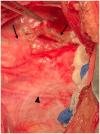
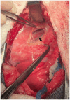
Open surgical treatment of idiopathic chylothorax via thoracic duct ligation and pericardiectomy requires a lengthy procedure with two thoracotomy incisions. The objectives of this report were to describe an approach for thoracic duct ligation and pericardiectomy via a single thoracotomy at the left fourth intercostal space and to describe the clinical outcome in two dogs with idiopathic chylothorax. Dogs were prospectively enrolled in a pilot study to evaluate the clinical efficacy of thoracic duct ligation at the left fourth intercostal space, combined with subphrenic pericardiectomy performed through the same incision. Dogs had a preoperative CT lymphangiogram to evaluate the anatomy of the thoracic duct and its branching pattern prior to surgery. Recheck radiographs were performed every 2-4 weeks until effusion resolved. Pleural effusion became non-chylous by 5 days postoperatively. Pleural effusion volume decreased by day 5 postoperatively, allowing removal of thoracostomy tube and discharge from the hospital. Radiographically, effusion resolved within 6 weeks without a need for further drainage after discharge. Dogs remained symptom-free at last follow up (>11 months postoperatively). CT lymphangiograms were repeated >11 months postoperatively and revealed no recurrence of pleural effusion. No intraoperative or postoperative complications directly related to surgery were noted for either dog. Collateral lymphatic vessels were not identified on recheck CT lymphangiograms. The left fourth intercostal approach to thoracic duct ligation and pericardiectomy has potential to be a safe and effective alternative to an open approach requiring two lateral thoracotomies. Further investigation of this approach using open or minimally invasive techniques is warranted.
Keywords: fourth intercostal space; idiopathic chylothorax; pericardiectomy; pleural effusion; thoracic duct ligation.
Copyright © 2024 Price and Mathews.
Conflict of interest statement
The authors declare that the research was conducted in the absence of any commercial or financial relationships that could be construed as a potential conflict of interest.
Figures


Similar articles
-
Evaluation of thoracic duct ligation and unilateral subphrenic pericardiectomy via a left fourth intercostal approach in normal canine cadavers.Vet Surg. 2024 Apr;53(3):437-446. doi: 10.1111/vsu.14060. Epub 2023 Dec 11. Vet Surg. 2024. PMID: 38078621 Review.
-
Triple-combination surgery with thoracic duct ligation, partial pericardiectomy, and cisterna chyli ablation for treatment of canine idiopathic chylothorax.J Vet Med Sci. 2022 Aug 1;84(8):1079-1083. doi: 10.1292/jvms.22-0043. Epub 2022 Jun 8. J Vet Med Sci. 2022. PMID: 35675979 Free PMC article.
-
Efficacy of en bloc thoracic duct ligation in combination with pericardiectomy by video-assisted thoracoscopic surgery for canine idiopathic chylothorax.Vet Surg. 2020 Jun;49 Suppl 1:O102-O111. doi: 10.1111/vsu.13370. Epub 2019 Dec 27. Vet Surg. 2020. PMID: 31880337
-
Minimally invasive treatment of idiopathic chylothorax in dogs by thoracoscopic thoracic duct ligation and subphrenic pericardiectomy: 6 cases (2007-2010).J Am Vet Med Assoc. 2012 Oct 1;241(7):904-9. doi: 10.2460/javma.241.7.904. J Am Vet Med Assoc. 2012. PMID: 23013503
-
Idiopathic chylothorax in dogs and cats: nonsurgical and surgical management.Compend Contin Educ Vet. 2012 Aug;34(8):E3. Compend Contin Educ Vet. 2012. PMID: 22935991 Review.
References
LinkOut - more resources
Full Text Sources

