. 2024 Sep 24;9(12):3580-3591.
doi: 10.1016/j.ekir.2024.09.013. eCollection 2024 Dec.

CFAP47 is Implicated in X-Linked Polycystic Kidney Disease

Collaborators, Affiliations

CFAP47 is Implicated in X-Linked Polycystic Kidney Disease

Takayasu Mori et al. Kidney Int Rep. .

Abstract

Introduction: Autosomal dominant polycystic kidney disease (ADPKD) is a well-described condition in which approximately 80% of all cases have a genetic explanation; and among sporadic cases without a family history, the genetic bases remain unclear in approximately 30% of cases. This study aimed to identify genes associated with polycystic kidney disease (PKD) in patients with sporadic cystic kidney disease in which a clear genetic change was not identified in established genes.

Methods: A next-generation sequencing panel analyzed known genes related to kidney cysts in 118 sporadic cases, followed by whole-genome sequencing (WGS) on 47 unrelated individuals without identified candidate variants. Immunohistology examination was then conducted on both human kidney tissue and kidneys from CFAP47-/Y mice.

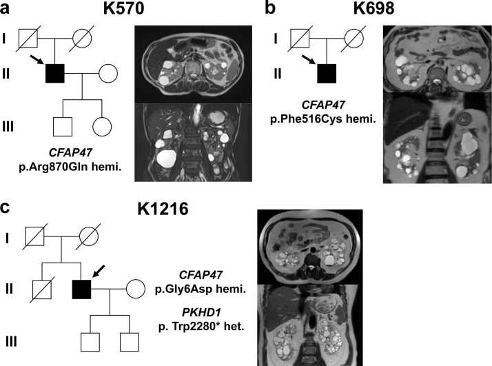
Results: Three male patients were found to have rare missense variants in the X-linked gene cilia and flagella-associated protein 47 (CFAP47), none of whom had a family history of the condition. CFAP47 was expressed in primary cilia of human kidney tubules, and knockout (KO) mice exhibited vacuolation of tubular cells and tubular dilation, providing evidence that CFAP47 is a causative gene involved in cyst formation.

Conclusion: This discovery of CFAP47 as a newly identified gene associated with PKD, displaying X-linked inheritance, emphasizes the need for further cases to understand the role of CFAP47 in PKD.

Keywords: CFAP47; X-linked; polycystic kidney disease; sporadic.


Figures

None
Graphical abstract
Figure 1
Figure 1
Summary of genetic analysis for a cohort of 118 cases with sporadic PKD. Targeted next generation sequencing panel analysis covering cystic kidney disease-related genes was followed by targeted long-read sequencing (T-LRS) and whole-genome sequencing (WGS). This approach identified CFAP47 mutations in 3 cases, constituting 2.5% of the total cohort. #The proportion within the 49 unsolved cases. PKD, polycystic kidney disease.
Figure 2
Figure 2
Family tree and MRI of polycystic kidneys. Family trees, genetic mutations, and abdominal MRI images of K570 (case 1), K698 (case 2), and K1216 (case 3) are shown in Panels A, B, and C, respectively. MRI, magnetic resonance imaging.
Figure 3
Figure 3
Evaluation of CFAP47 expression sites in human kidney tubules and kidney morphology of CFAP47−/Y mice. (a) Human kidney normal tubules coimmunostained with acetylated alpha-tubulin antibody (red) and CFAP47 antibody (green); (b) CFAP47+/Y AND CFAP47−/Y mouse kidney macro-morphology images; (c) kidney volume comparison using the ellipsoid formula: (length × width × [π/6]) (d) Low and high magnification images with hematoxylin and eosin stain.
Figure 4
Figure 4
Evaluation of vacuolated cell localization, and primary cilia length in CFAP47−/Y mice. (a) Paraffin sections of Cfap47−/Y mouse kidneys were stained using an enzyme antibody method with proximal tubule marker (AQP1) and distal tubule marker (NCC [SLC12A3]). (b) Fluorescent immunostaining was performed using acetylated alpha-tubulin antibody (red) to detect primary cilia in the proximal tubules of kidneys from CFAP47+/Y AND CFAP47−/Y mice. Arrowheads indicate primary cilia. (c) The length of primary cilia stained in (b) was compared between CFAP47+/Y and CFAP47−/Y and presented as a box plot. DCT, distal convoluted tubule; PT, proximal tubule.

Update of

References

    1. Rossetti S., Consugar M.B., Chapman A.B., et al. Comprehensive molecular diagnostics in autosomal dominant polycystic kidney disease. J Am Soc Nephrol. 2007;18:2143–2160. doi: 10.1681/ASN.2006121387. - DOI - PubMed
    1. Mallawaarachchi A.C., Lundie B., Hort Y., et al. Genomic diagnostics in polycystic kidney disease: an assessment of real-world use of whole-genome sequencing. Eur J Hum Genet. 2021;29:760–770. doi: 10.1038/s41431-020-00796-4. - DOI - PMC - PubMed
    1. Chang A.R., Moore B.S., Luo J.Z., et al. Exome sequencing of a clinical population for autosomal dominant polycystic kidney disease. JAMA. 2022;328:2412–2421. doi: 10.1001/jama.2022.22847. - DOI - PMC - PubMed
    1. Yu C.C., Lee A.F., Kohl S., et al. PKD2 founder mutation is the most common mutation of polycystic kidney disease in Taiwan. NPJ Genom Med. 2022;7:40. doi: 10.1038/s41525-022-00309-w. - DOI - PMC - PubMed
    1. Porath B., Gainullin V.G., Cornec-Le Gall E., et al. Mutations in GANAB, encoding the glucosidase IIα subunit, cause autosomal-dominant polycystic kidney and liver disease. Am J Hum Genet. 2016;98:1193–1207. doi: 10.1016/j.ajhg.2016.05.004. - DOI - PMC - PubMed

LinkOut - more resources