. 2024 Feb 29;5(1):119-137.
doi: 10.20517/evcna.2023.55. eCollection 2024.

A comparative analysis of small extracellular vesicle (sEV) micro-RNA (miRNA) isolation and sequencing procedures in blood plasma samples

Affiliations

A comparative analysis of small extracellular vesicle (sEV) micro-RNA (miRNA) isolation and sequencing procedures in blood plasma samples

Pevindu Abeysinghe et al. Extracell Vesicles Circ Nucl Acids. .

Abstract

Aims: Analysis of miRNA (18-23nt) encapsulated in small extracellular vesicles (sEVs) (diameter ~30-200 nm) is critical in understanding the diagnostic and therapeutic value of sEV miRNA. However, various sEV enrichment techniques yield different quantities and qualities of sEV miRNA. Here, we compare the efficacy of three sEV isolation techniques in four combinations for miRNA next-generation sequencing.

Methods: Blood plasma from four Holstein-Friesian dairy cows (Bos taurus) (n = 4) with similar genetic traits and physical characteristics were pooled to isolate sEV. Ultracentrifugation (UC) (100,000 × g, 2 h at 4 °C), size-exclusion chromatography (SEC) and ultrafiltration (UF) were used to design four groups of sEV isolations (UC+SEC, SEC+UC, SEC+UF and UC+SEC+UF). sEV miRNAs were isolated using a combination of TRIzol, Chloroform and miRNeasy mini kit (n = 4/each), later sequenced utilizing Novaseq S1 platform (single-end 100 bp sequencing).

Results: All four sEV methods yielded > 1,700 miRNAs and sEV miRNAs demonstrated a clear separation from control blood plasma circulating miRNA (PCA analysis). MiR-381-3p, miR-23-3p, and miR-18b-3p are among the 25 miRNAs unique to sEV, indicating potential sEV-specific miRNA markers. Further, those 25 miRNAs mostly regulate immune-related functions, indicating the value of sEV miRNA cargo in immunology.

Conclusion: The four sEV miRNA isolation methods employed in this study are valid techniques. The choice of method depends on the research question and study design. If purity is of concern, the UC+SEC method resulted in the best particles/µg protein ratio, which is often used as an indication of sample purity. These results could eventually establish sEV miRNAs as effective diagnostic and therapeutic tools of immunology.

Keywords: Small extracellular vesicles; exosomes; immune function; miRNA; next-generation sequencing; size exclusion chromatography; ultracentrifugation; ultrafiltration.


Conflict of interest statement

All authors declared that there are no conflicts of interest.

Figures

Figure 1
Figure 1
Schematic diagram of sEV isolation methods (Created with BioRender.com).
Figure 2
Figure 2
Schematic diagram of optimized miRNA isolation methodology (Created with BioRender.com).
Figure 3
Figure 3
Characterization of sEV using NTA, western blot and TEM. (A) box plot (line at mean) of the number of particles per µg of total protein present per mL of sEV sample for the four different purification methods (*** P = 0.0008, **P = 0.0015, *P = 0.017). Three biological replicates were collected at the end of each isolation method for NTA; (B) relative abundance of exosome/EV membrane proteins (CD81, CD9, and Flot-1) and non-exosome protein marker (BSA) in sEV samples isolated from four different methodologies. Three biological replicates were pooled to create a representative sample, which was then utilized for three technical replicates (3 lanes in the Western Blot gel). Positive control is bovine plasma; (C) TEM images depict the spherical and cup-shaped sEV vesicles with appropriate size (~30-200 nm); (D) representation of original NTA images demonstrates that the EVs isolated from each method predominantly fall approximately between 30-200 nm in size.
Figure 4
Figure 4
sEV miRNA characterization and next-generation sequencing demonstrated all four sEV miRNA isolation methodological approaches were successful (four technical replicates were utilized (n = 4) for each of the four isolation methods). (A) Quality control (QC) results of sEV miRNA samples quantified as pg/mL of miRNA concentration; (B) Summary of next-generation sequencing (NGS): different types of RNA read counts identified; (C) Total number of bovine (Bos taurus) miRNA counts identified; (D) the Venn diagram of shared and unique miRNAs in each method compared to the blood plasma control; (E) the principal components analysis (PCA) plot illustrates all sEV miRNA samples cluster together and away from the blood plasma-derived miRNA control. UC: Ultracentrifugation; SEC: Size exclusion chromatography; UF: Ultrafiltration.
Figure 5
Figure 5
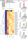
Unique miRNAs present in sEV compared to blood plasma. (A) Heatmap of DE miRNAs; Expression of miRNAs. Box and whiskers (minimum to maximum) plots of (B) miR-381-3p; (C) miR-101-1-5p; (D) miR-144-3p; (E) miR-23b-3p. BP: blood plasma; UC: ultracentrifugation; SEC: size-exclusion chromatography; UF: ultrafiltration; DE: differential expression.
Figure 6
Figure 6
DE miRNAs present in blood plasma compared to sEV. (A) Heatmap of top 25 DE miRNAs. Expression of miRNAs. Box and whiskers (minimum to maximum) plots of (B) miR-2285ah-3p; (C) miR-214-3p; (D) miR-10020-5p; (E) miR-12024-5p. BP: Blood plasma; UC: Ultracentrifugation; SEC: Size-exclusion chromatography; UF: Ultrafiltration; DE: differential expression.
Figure 7
Figure 7
Gene target prediction and Gene Ontology (GO) enrichment analysis for miRNA uniquely present in sEV. (A) gene targets for 25 miRNA uniquely present in sEV were identified using miRNET, targetScan, and miRWalk miRNA target prediction tools and identified 95 shared/overlapping genes; (B) pathway analysis identified 95 genes that regulate mostly immune and inflammatory function (highlighted as exploded portions of the pie chart). This indicates 25 miRNA unique to sEV relates to immune function.
Figure 8
Figure 8
Comparative analysis of cellular component GO enrichment profiles for miRNA expression across four isolation methods.

Similar articles

Cited by

References

    1. Min L, Zhu S, Chen L, et al. Evaluation of circulating small extracellular vesicles derived miRNAs as biomarkers of early colon cancer: a comparison with plasma total miRNAs. J Extracell Vesicles. 2019;8:1643670. doi: 10.1080/20013078.2019.1643670. - DOI - PMC - PubMed
    1. Rajkumar AP, Hye A, Lange J, et al. Next-generation RNA-sequencing of serum small extracellular vesicles discovers potential diagnostic biomarkers for dementia with lewy bodies. Am J Geriatr Psychiatry. 2021;29:573–84. doi: 10.1016/j.jagp.2020.10.012. - DOI - PubMed
    1. Jiang YF, Wei SN, Geng N, et al. Evaluation of circulating small extracellular vesicle-derived miRNAs as diagnostic biomarkers for differentiating between different pathological types of early lung cancer. Sci Rep. 2022;12:17201. doi: 10.1038/s41598-022-22194-0. - DOI - PMC - PubMed
    1. Buschmann D, Kirchner B, Hermann S, et al. Evaluation of serum extracellular vesicle isolation methods for profiling miRNAs by next-generation sequencing. J Extracell Vesicles. 2018;7:1481321. doi: 10.1080/20013078.2018.1481321. - DOI - PMC - PubMed
    1. Grunt M, Failla AV, Stevic I, Hillebrand T, Schwarzenbach H. A novel assay for exosomal and cell-free miRNA isolation and quantification. RNA Biol. 2020;17:425–40. doi: 10.1080/15476286.2020.1721204. - DOI - PMC - PubMed

LinkOut - more resources