

Myocardial perfusion imaging in a case of cardiac strangulation: A novel approach to detect pacemaker-induced ischemia
- PMID: 39698428
- PMCID: PMC11651413
- DOI: 10.4103/apc.apc_125_24
Myocardial perfusion imaging in a case of cardiac strangulation: A novel approach to detect pacemaker-induced ischemia
Abstract
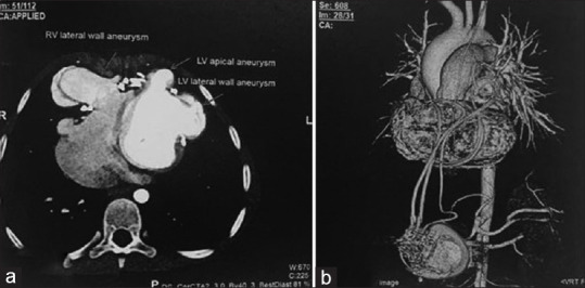
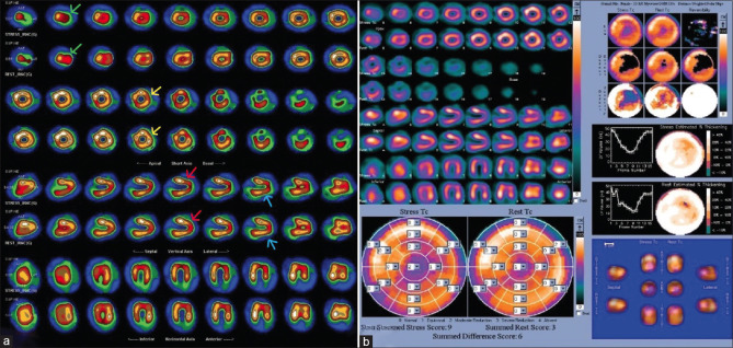
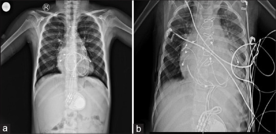
Cardiac strangulation is a rare phenomenon in children following epicardial pacemaker implantation, caused by compression of the heart and great vessels by the epicardial pacemaker wires. We report a rare case of cardiac strangulation presenting after 8 years of epicardial pacemaker implantation. On routine follow-up, computed tomography angiography showed significant extrinsic compression of the mid-left anterior descending (LAD) artery by the epicardial pacing wire. She was referred to our department for myocardial perfusion imaging (MPI), which showed significant inducible ischemia in the LAD territory. Following this, she underwent a successful pacemaker reimplantation. MPI can, thus, act as a good tool to assess the functional significance of the compression caused by strangulation of the heart by pacemaker leads in asymptomatic patients.
Keywords: Congenital heart block; myocardial ischemia; pacing in children.
Copyright: © 2024 Annals of Pediatric Cardiology.
Conflict of interest statement
There are no conflicts of interest.
Figures
References
-
- Carreras EM, Duncan WJ, Djurdjev O, Campbell AI. Cardiac strangulation following epicardial pacemaker implantation: A rare pediatric complication. J Thorac Cardiovasc Surg. 2015;149:522–7. - PubMed
-
- Morrison ML, Karayiannis S, Speggiorin S, Sands AJ, Rosenthal E. Cardiac strangulation after epicardial pacing: The importance of non-specific symptoms and a low index of suspicion. Cardiol Young. 2021;31:159–62. - PubMed
-
- Mah DY, Prakash A, Porras D, Fynn-Thompson F, DeWitt ES, Banka P. Coronary artery compression from epicardial leads: More common than we think. Heart Rhythm. 2018;15:1439–47. - PubMed
Publication types
LinkOut - more resources
Full Text Sources