Review
. 2024 Jun 18;5(2):297-328.
doi: 10.20517/evcna.2024.21. eCollection 2024.

Cargo exchange between human and bacterial extracellular vesicles in gestational tissues: a new paradigm in communication and immune development

Affiliations
Review

Cargo exchange between human and bacterial extracellular vesicles in gestational tissues: a new paradigm in communication and immune development

Emmanuel Amabebe et al. Extracell Vesicles Circ Nucl Acids. .

Abstract

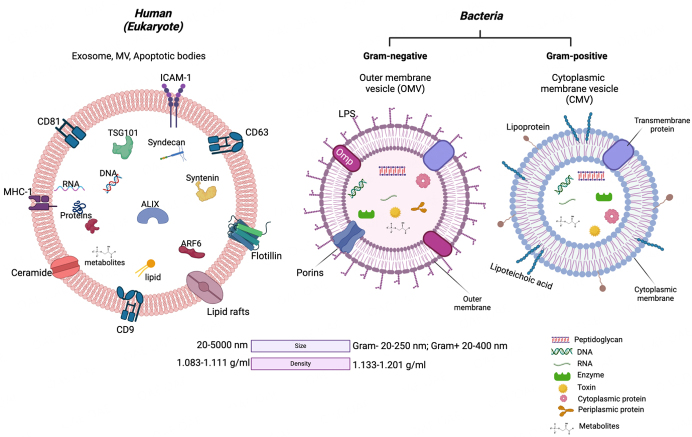
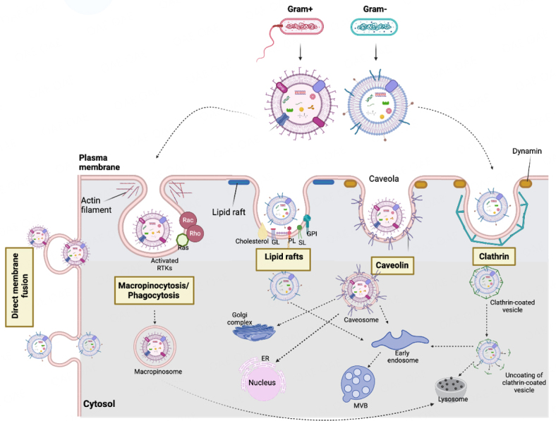
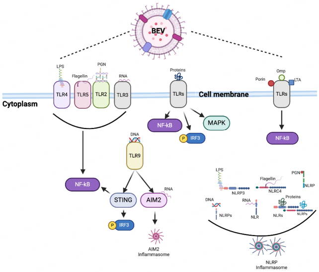
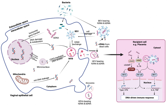
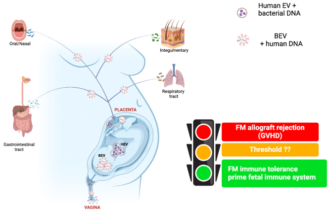
Host-bacteria and bacteria-bacteria interactions can be facilitated by extracellular vesicles (EVs) secreted by both human and bacterial cells. Human and bacterial EVs (BEVs) propagate and transfer immunogenic cargos that may elicit immune responses in nearby or distant recipient cells/tissues. Hence, direct colonization of tissues by bacterial cells is not required for immunogenic stimulation. This phenomenon is important in the feto-maternal interface, where optimum tolerance between the mother and fetus is required for a successful pregnancy. Though the intrauterine cavity is widely considered sterile, BEVs from diverse sources have been identified in the placenta and amniotic cavity. These BEVs can be internalized by human cells, which may help them evade host immune surveillance. Though it appears logical, whether bacterial cells internalize human EVs or human EV cargo is yet to be determined. However, the presence of BEVs in placental tissues or amniotic cavity is believed to trigger a low-grade immune response that primes the fetal immune system for ex-utero survival, but is insufficient to disrupt the progression of pregnancy or cause immune intolerance required for adverse pregnancy events. Nevertheless, the exchange of bioactive cargos between human and BEVs, and the mechanical underpinnings and health implications of such interactions, especially during pregnancy, are still understudied. Therefore, while focusing on the feto-maternal interface, we discussed how human cells take up BEVs and whether bacterial cells take up human EVs or their cargo, the exchange of cargos between human and BEVs, host cell (feto-maternal) inflammatory responses to BEV immunogenic stimulation, and associations of these interactions with fetal immune priming and adverse reproductive outcomes such as preeclampsia and preterm birth.

Keywords: Extracellular vesicles; fetal membranes; feto-maternal interface; immune priming; outer membrane vesicles; placenta.


Conflict of interest statement

All authors declared that there are no conflicts of interest.

Figures

Figure 1
Figure 1
Comparison of human and bacterial-derived extracellular vesicles. ARF6: ADP ribosylation factor 6; ESCRT proteins (TSG101, ALIX); ICAM-1: intercellular adhesion molecule 1; LPS: lipopolysaccharide; Omp: outer membrane protein; MHC-1: major histocompatibility complex-1; Nuclei acids (DNA, RNA); Tetraspanins (CD9, CD63, CD81). Density data[130]. Created with BioRender.com.
Figure 2
Figure 2
Mechanisms of uptake of bacterial extracellular vesicles by human (host) cells. BEVs enter human cells by macropinocytosis/phagocytosis, clathrin- and caveolin-mediated endocytosis, lipid rafts, and direct fusion with the plasma membrane. ER: endoplasmic reticulum; MVB: multivesicular bodies; GL: glycolipid; GPI: GPI-anchored proteins; PL: phospholipid; RTK: receptor tyrosine kinases; SL: sphingolipid. Created with BioRender.com.
Figure 3
Figure 3
Bacterial extracellular vesicle (BEV) pathogen-associated molecular patterns (PAMPs) recognize surface membrane and cytoplasmic host cell pattern recognition receptors and activate downstream inflammatory signaling pathways. Omp, LTA, and porin only bind to TLRs on the cell membrane surface. At the same time, both surface membrane and cytoplasmic receptors recognize the other PAMPs. AIM2: absent in melanoma 2; IRF: interferon regulatory transcription factor; LPS: lipopolysaccharide; LTA: lipoteichoic acid; MAPK: mitogen-associated protein kinase; NF-κB: nuclear factor kappa B; NLR: nucleotide-binding oligomerization domain-like receptor; NLRP: NOD-like receptor thermal protein domain-associated protein; Omp: outer membrane protein; PGN: peptidoglycan; STING: stimulator of interferon genes; TLR: toll-like receptor. Created with BioRender.com.
Figure 4
Figure 4
DNA-driven immune response mediated by DNA exchange between human and bacterial extracellular vesicles. Human/bacterial genomic/mitochondrial DNA carried by EVs can be delivered to cells as damage- (DAMP) or pathogen-associated molecular pattern (PAMP). EV-DNA can activate inflammatory pathways through cytosolic DNA receptors such as cGAS (cyclic GMP-AMP) and AIM2 (absent in melanoma 2 or interferon-inducible protein). Activation of the cGAS/STING pathway causes downstream release of type I interferons (IFN-α, IFN-β, IFN-ε). Activation of AIM2 produces interleukins and tumor necrosis factor (TNF). This may manifest as a subclinical sterile inflammatory response in gestational tissues such as the placenta and amniochorion. bDNA: bacterial DNA; BEV: bacterial extracellular vesicles; cfDNA: cell-free DNA; DNA: human DNA; HEV: human extracellular vesicles; IRF: interferon regulatory factor; mtDNA: mitochondrial DNA; MVB: multivesicular bodies; NF-κB: nuclear factor-kappa B; STING: stimulator of interferon genes. Created with BioRender.com.
Figure 5
Figure 5
Extracellular vesicle-mediated feto-maternal immune tolerance. The feto-maternal immune tolerance that maintains normal pregnancy is partly mediated by BEVs from various microbiotas. Maternal BEVs can also carry DAMPs/PAMPs to the feto-maternal interface (placenta tissues) and trigger adverse immune responses that may manifest as feto-maternal allograft rejection in the form of graft-versus-host disease (GVHD). However, the threshold at which such a switch occurs, and the mechanisms involved are yet to be determined. Conversely, pathogenic bacteria can shed BEVs that can cause inflammatory responses often associated with adverse pregnancy conditions. BEV: bacterial extracellular vesicle; DAMPs/PAMPs: damage- and pathogen-associated molecular patterns; FM: feto-maternal; HEV: human extracellular vesicle. Created with BioRender.com.

Similar articles

Cited by

References

    1. Gill S, Catchpole R, Forterre P. Extracellular membrane vesicles in the three domains of life and beyond. FEMS Microbiol Rev. 2019;43:273–303. doi: 10.1093/femsre/fuy042. - DOI - PMC - PubMed
    1. Ñahui Palomino RA, Vanpouille C, Costantini PE, Margolis L. Microbiota-host communications: bacterial extracellular vesicles as a common language. PLoS Pathog. 2021;17:e1009508. doi: 10.1371/journal.ppat.1009508. - DOI - PMC - PubMed
    1. Welsh JA, Goberdhan DCI, O'Driscoll L, et al. MISEV Consortium. Minimal information for studies of extracellular vesicles (MISEV2023): from basic to advanced approaches. J Extracell Vesicles. 2024;13:e12404. doi: 10.1002/jev2.12404. - DOI - PMC - PubMed
    1. Bose S, Aggarwal S, Singh DV, Acharya N. Extracellular vesicles: an emerging platform in gram-positive bacteria. Microb Cell. 2020;7:312–22. doi: 10.15698/mic2020.12.737. - DOI - PMC - PubMed
    1. Sáez T, de Vos P, Sobrevia L, Faas MM. Is there a role for exosomes in foetoplacental endothelial dysfunction in gestational diabetes mellitus? Placenta. 2018;61:48–54. doi: 10.1016/j.placenta.2017.11.007. - DOI - PubMed

LinkOut - more resources