

doi: 10.21037/qims-24-949.
Epub 2024 Nov 18.
Symptomatic air embolism after computed tomography-guided four-hook needle localization of a pulmonary nodule: a case description
Affiliations
- PMID: 39698648
- PMCID: PMC11652006
- DOI: 10.21037/qims-24-949
Item in Clipboard
Symptomatic air embolism after computed tomography-guided four-hook needle localization of a pulmonary nodule: a case description
Quant Imaging Med Surg.
.
No abstract available
Conflict of interest statement
Conflict of Interest: All authors have completed the ICMJE uniform disclosure form (available at https://qims.amegroups.com/article/view/10.21037/qims-24-949/coif). The authors have no conflicts of interest to declare.
Figures
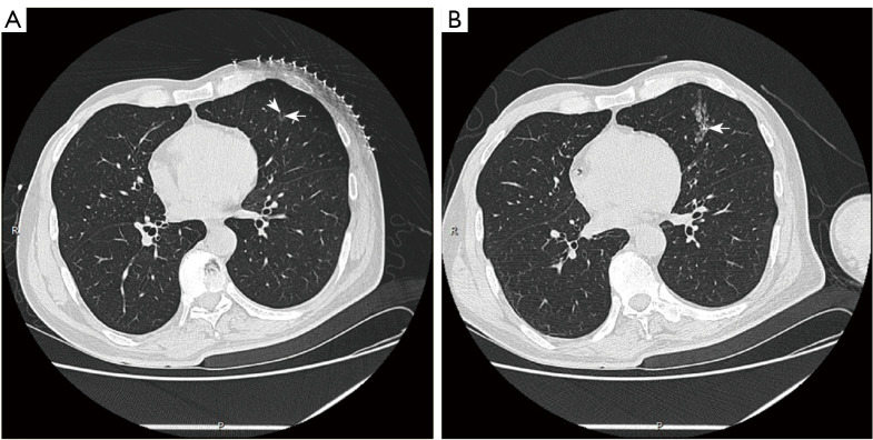
A 62-year-old female with a ground-glass nodule in the left upper lobe underwent CT-guided localization with a four-hook needle. (A) The lesion (arrow) observed on an axial CT image. (B) Postlocalization CT showed the location of the released anchor claw (arrow). The bronchial passage was visible within the pulmonary nodule, and small pulmonary veins could be seen in front of the nodule (arrowhead in A). CT, computed tomography.
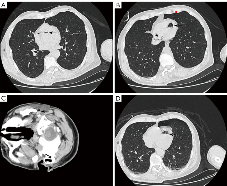
The chest and brain CT revealed systemic air embolism after four-hook needle localization. (A) An air-fluid level was observed at the aortic root (arrow). (B) A mass of air was visible in the left ventricle (arrow), the right coronary artery (arrowhead), and the left internal mammary artery (red arrow). (C) Air embolism was also detected in the left external carotid artery (arrow). (D) Forty minutes after the initial brain CT scan, a second CT scan showed the disappearance of air in the aortic root, right coronary artery, left internal mammary artery, and left external carotid artery; moreover, the volume of air in the left ventricle was significantly reduced (arrow). CT, computed tomography.
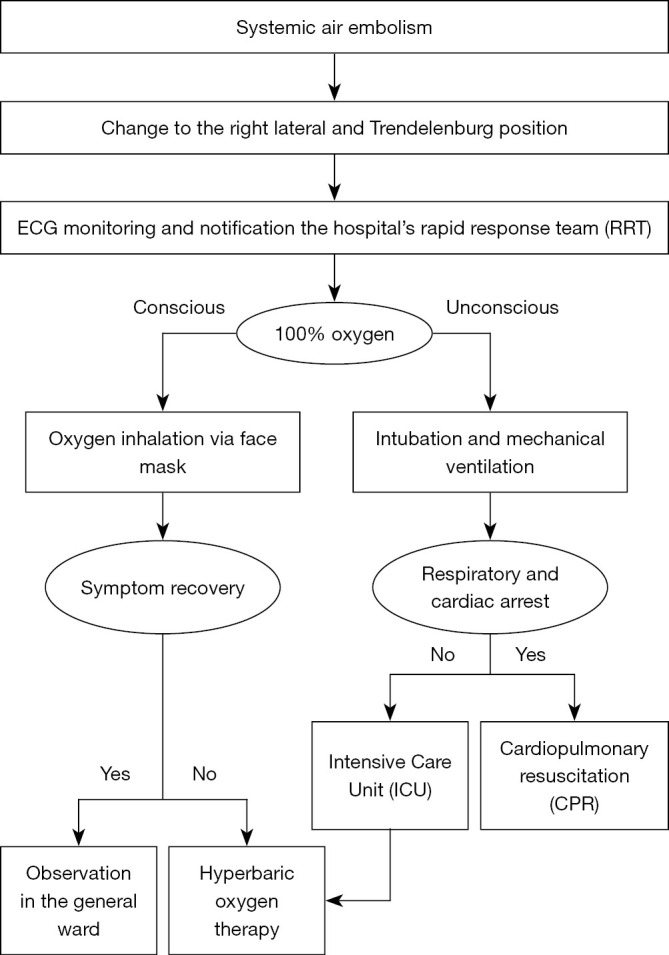
Flowchart depicting the management of systemic air embolism after the lung localization procedure.
References
LinkOut - more resources
Full Text Sources