

A literature review of artificial intelligence (AI) for medical image segmentation: from AI and explainable AI to trustworthy AI
- PMID: 39698664
- PMCID: PMC11651983
- DOI: 10.21037/qims-24-723
A literature review of artificial intelligence (AI) for medical image segmentation: from AI and explainable AI to trustworthy AI
Abstract
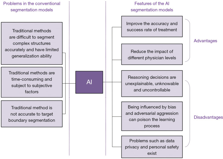
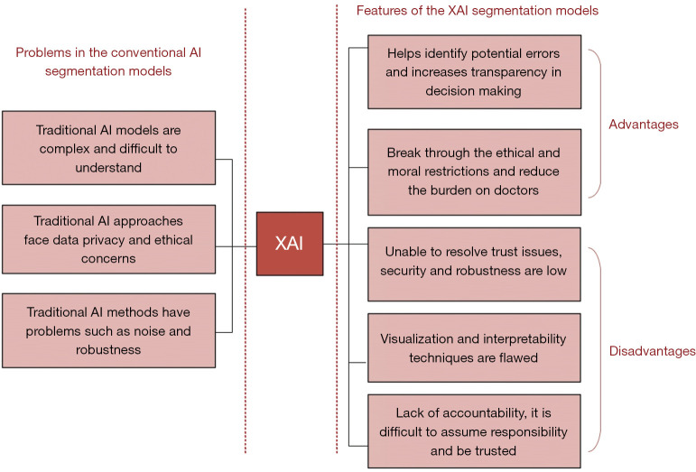
Background and objective: Medical image segmentation is a vital aspect of medical image processing, allowing healthcare professionals to conduct precise and comprehensive lesion analyses. Traditional segmentation methods are often labor intensive and influenced by the subjectivity of individual physicians. The advent of artificial intelligence (AI) has transformed this field by reducing the workload of physicians, and improving the accuracy and efficiency of disease diagnosis. However, conventional AI techniques are not without challenges. Issues such as inexplicability, uncontrollable decision-making processes, and unpredictability can lead to confusion and uncertainty in clinical decision-making. This review explores the evolution of AI in medical image segmentation, focusing on the development and impact of explainable AI (XAI) and trustworthy AI (TAI).
Methods: This review synthesizes existing literature on traditional segmentation methods, AI-based approaches, and the transition from conventional AI to XAI and TAI. The review highlights the key principles and advancements in XAI that aim to address the shortcomings of conventional AI by enhancing transparency and interpretability. It further examines how TAI builds on XAI to improve the reliability, safety, and accountability of AI systems in medical image segmentation.
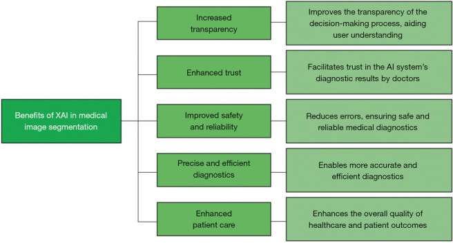
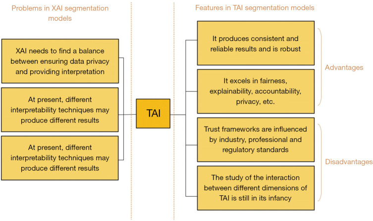
Key content and findings: XAI has emerged as a solution to the limitations of conventional AI by providing greater transparency and interpretability, allowing healthcare professionals to better understand and trust AI-driven decisions. However, XAI itself faces challenges, including those related to safety, robustness, and value alignment. TAI has been developed to overcome these challenges, offering a more reliable framework for AI applications in medical image segmentation. By integrating the principles of XAI with enhanced safety and dependability, TAI addresses the critical need for TAI systems in clinical settings.
Conclusions: TAI presents a promising future for medical image segmentation, combining the benefits of AI with improved reliability and safety. Thus, TAI is a more viable and dependable option for healthcare applications, and could ultimately lead to better clinical outcomes for patients, and advance the field of medical image processing.
Keywords: Medical image segmentation; artificial intelligence (AI); explainable AI (XAI); trustworthy AI (TAI).
2024 AME Publishing Company. All rights reserved.
Conflict of interest statement
Conflicts of Interest: All authors have completed the ICMJE uniform disclosure form (available at https://qims.amegroups.com/article/view/10.21037/qims-24-723/coif). The authors have no conflicts of interest to declare.
Figures
References
-
- Lee LK, Liew SC, Thong WJ. A review of image segmentation methodologies in medical image. Adv Comput Commun Eng Technol 2015;1069-80.
-
- Chen A, Zhu L, Zang H, Ding Z, Zhan S. Computer-aided diagnosis and decision-making system for medical data analysis: a case study on prostate MR images. J Manag Sci Eng 2019;4:266-78.
-
- Abdel-Basset M, Chang V, Mohamed R. A novel equilibrium optimization algorithm for multi-thresholding image segmentation problems. Neural Comput Appl 2021;33:10685-718.
-
- Yang D, Xu Z, Li W, Myronenko A, Roth HR, Harmon S, Xu S, Turkbey B, Turkbey E, Wang X, Zhu W, Carrafiello G, Patella F, Cariati M, Obinata H, Mori H, Tamura K, An P, Wood BJ, Xu D. Federated semi-supervised learning for COVID region segmentation in chest CT using multi-national data from China, Italy, Japan. Med Image Anal 2021;70:101992. 10.1016/j.media.2021.101992 - DOI - PMC - PubMed
Publication types
LinkOut - more resources
Full Text Sources