Review
. 2024 Dec 5;14(12):9589-9599.
doi: 10.21037/qims-24-34. Epub 2024 Oct 18.

Role of high-resolution magnetic resonance imaging in preoperative tumor-node-metastasis staging evaluation of esophageal cancer: a narrative review

Affiliations
Review

Role of high-resolution magnetic resonance imaging in preoperative tumor-node-metastasis staging evaluation of esophageal cancer: a narrative review

Yingyu Lin et al. Quant Imaging Med Surg. .

Abstract

Background and objective: Esophageal cancer (EC) is an aggressive disease characterized by high mortality rates and a propensity for locoregional or distant recurrence. The treatment strategies and prognostic estimation for EC depend on accurate pre-treatment tumor-node-metastasis (TNM) staging. The objective of this review was to illustrate the role of various imaging modalities in achieving accurate preoperative TNM staging of EC, with a particular focus on the utilization of advanced high-resolution magnetic resonance imaging (MRI) sequences for T classification, which have shown promise in enhancing the delineation of tumor depth and extent.

Methods: A comprehensive literature search was conducted in PubMed and Web of Science databases. The studies on imaging in preoperative TNM staging of EC published in English from inception of these databases to December 31, 2022 were reviewed.

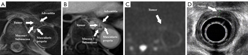
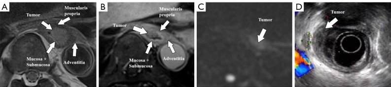
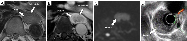
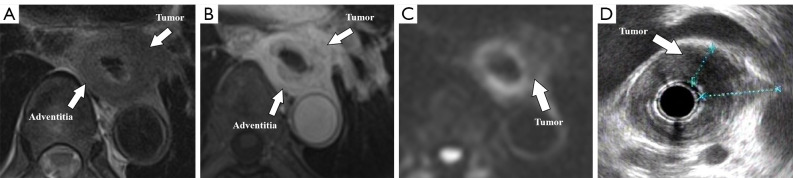
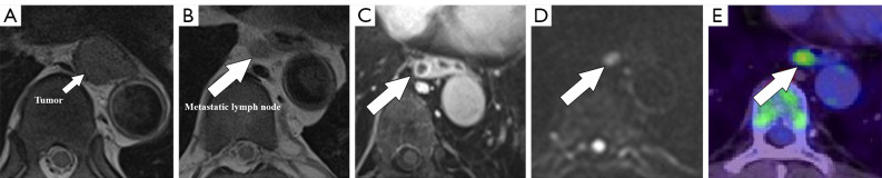
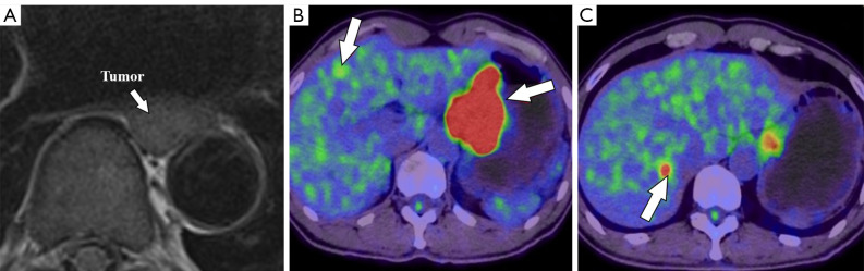
Key content and findings: The review highlights the distinct advantages and inherent limitations of different imaging modalities for the preoperative staging of EC. Endoscopic ultrasound (EUS) provides real-time, high-resolution imaging of the esophageal wall but is operator-dependent. Computed tomography (CT) is widely available and non-invasive, but it may lack sensitivity for early T-stage identification. Positron emission tomography (PET)/CT offers accurate assessment of distant metastasis but has limited value in the evaluation of early-stage tumors. With improved techniques, MRI is particularly useful for visualization of tumor infiltration and the surrounding anatomical structures, gaining prominence in preoperative staging of EC.

Conclusions: Various imaging modalities including EUS, CT, PET/CT, and MRI should be applied as complementary methods for preoperative TNM staging of EC. Notably, high-resolution MRI can overcome motion-related artifacts and provide high-quality images, which may play a more important role in the management of EC in the future.

Keywords: Esophageal cancer (EC); magnetic resonance imaging (MRI); preoperative assessment; tumor-node-metastasis staging (TNM staging).


Conflict of interest statement

Conflicts of Interest: All authors have completed the ICMJE uniform disclosure form (available at https://qims.amegroups.com/article/view/10.21037/qims-24-34/coif). The authors have no conflicts of interest to declare.

Figures

Figure 1
Figure 1
A 49-year-old male patient with T1 esophageal cancer: (A) T2WI BLADE, (B) enhanced T1WI StarVIBE, (C) DWI (b =800), and (D) EUS. The tumor is located in the mucosa and submucosa and protrudes into the lumen without muscularis propria invasion. T2WI, T2-weighted imaging; T1WI, T1-weighted imaging; StarVIBE, stack-of-stars volume interpolated breath-hold examination; DWI, diffusion-weighted imaging; EUS, endoscopic ultrasound.
Figure 2
Figure 2
A 68-year-old male patient with T2 esophageal cancer: (A) T2WI BLADE, (B) enhanced T1WI StarVIBE, (C) DWI (b =800), and (D) EUS. The tumor breaks through the submucosa and is limited to the muscularis propria. T2WI, T2-weighted imaging; T1WI, T1-weighted imaging; StarVIBE, stack-of-stars volume interpolated breath-hold examination; DWI, diffusion-weighted imaging; EUS, endoscopic ultrasound.
Figure 3
Figure 3
A 70-year-old male patient with T3 esophageal cancer: (A) T2WI BLADE, (B) enhanced T1WI StarVIBE, (C) DWI (b =800), and (D) EUS. The tumor is confined to the adventitia. T2WI, T2-weighted imaging; T1WI, T1-weighted imaging; StarVIBE, stack-of-stars volume interpolated breath-hold examination; DWI, diffusion-weighted imaging; EUS, endoscopic ultrasound.
Figure 4
Figure 4
A 61-year-old male patient with T4 esophageal cancer: (A) T2WI BLADE, (B) enhanced T1WI StarVIBE, (C) DWI (b =800), and (D) EUS. The tumor invades the pleura. T2WI, T2-weighted imaging; T1WI, T1-weighted imaging; StarVIBE, stack-of-stars volume interpolated breath-hold examination; DWI, diffusion-weighted imaging; EUS, endoscopic ultrasound.
Figure 5
Figure 5
A 55-year-old male esophageal cancer patient with local lymph node metastasis: (A,B) T2WI BLADE, (C) enhanced T1WI StarVIBE, (D) DWI (b =800), and (E) PET/CT. Metastatic lymph node with ring enhancement (white arrow in C), restricted diffusion in DWI (b =800) (white arrow in D) and high 18F-FDG uptake (white arrow in E). T2WI, T2-weighted imaging; T1WI, T1-weighted imaging; StarVIBE, stack-of-stars volume interpolated breath-hold examination; DWI, diffusion-weighted imaging; PET/CT, positron emission tomography/computed tomography; 18F-FDG, 18F-fluorodeoxyglucose.
Figure 6
Figure 6
A 62-year-old male esophageal cancer patient with liver metastases: (A) T2WI BLADE and (B,C) PET/CT. The liver metastases exhibit high metabolism (white arrows in B and C). T2WI, T2-weighted imaging; PET/CT, positron emission tomography/computed tomography.

Similar articles

References

    1. Bray F, Laversanne M, Sung H, Ferlay J, Siegel RL, Soerjomataram I, Jemal A. Global cancer statistics 2022: GLOBOCAN estimates of incidence and mortality worldwide for 36 cancers in 185 countries. CA Cancer J Clin 2024;74:229-63. 10.3322/caac.21834 - DOI - PubMed
    1. An L, Zheng R, Zeng H, Zhang S, Chen R, Wang S, Sun K, Li L, Wei W, He J. The survival of esophageal cancer by subtype in China with comparison to the United States. Int J Cancer 2023;152:151-61. 10.1002/ijc.34232 - DOI - PubMed
    1. Ajani JA, D'Amico TA, Bentrem DJ, Cooke D, Corvera C, Das P, et al. Esophageal and Esophagogastric Junction Cancers, Version 2.2023, NCCN Clinical Practice Guidelines in Oncology. J Natl Compr Canc Netw 2023;21:393-422. 10.6004/jnccn.2023.0019 - DOI - PubMed
    1. Siewert JR, Ott K. Are squamous and adenocarcinomas of the esophagus the same disease? Semin Radiat Oncol 2007;17:38-44. 10.1016/j.semradonc.2006.09.007 - DOI - PubMed
    1. Obermannová R, Alsina M, Cervantes A, Leong T, Lordick F, Nilsson M, van Grieken NCT, Vogel A, Smyth EC, ESMO Guidelines Committee . Electronic address: clinicalguidelines@esmo. Oesophageal cancer: ESMO Clinical Practice Guideline for diagnosis, treatment and follow-up. Ann Oncol 2022;33:992-1004. 10.1016/j.annonc.2022.07.003 - DOI - PubMed

LinkOut - more resources