

Evaluation of neostriatum changes in Crohn's disease: a multimodal brain magnetic resonance imaging study
- PMID: 39698710
- PMCID: PMC11651968
- DOI: 10.21037/qims-23-1603
Evaluation of neostriatum changes in Crohn's disease: a multimodal brain magnetic resonance imaging study
Abstract
Background: Abnormalities of neostriatum have been reported to be implicated in Crohn's disease (CD). However, there are few systematic explorations. We aim to explore the changes that occur in the structure and function of the neostriatum and whether these changes are related to the clinical characteristics of CD.
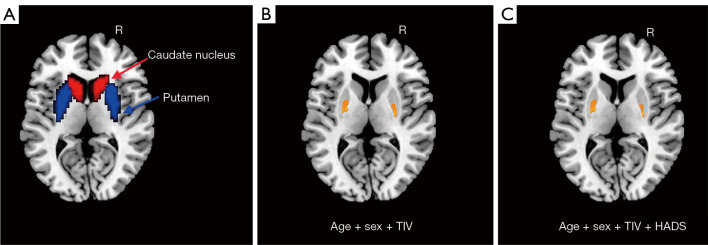
Methods: In this cross-sectional and prospective study, we enrolled 34 CD patients and 31 healthy controls (HCs) for analysis. We performed voxel-based morphometry (VBM) and seed-based functional connectivity (FC) to evaluate the structural and functional changes in the neostriatum. Correlation analysis was used to evaluate the possible relationships between clinical characteristics and neuroimaging findings.
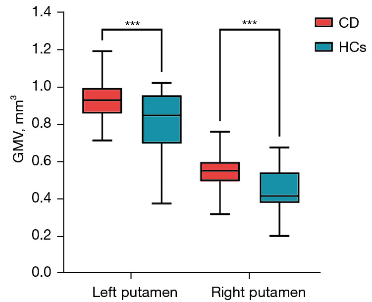
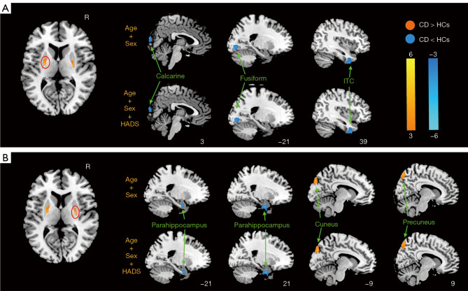
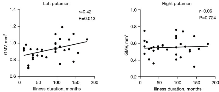
Results: CD patients had significantly increased gray matter volume (GMV) in the bilateral putamen compared with HCs. The results showed that CD patients had significantly decreased FC related to the putamen-calcarine cortex, putamen-fusiform gyrus, putamen-inferior temporal cortex (ITC), putamen-parahippocampus, and increased FC associated with the putamen-cuneus/precuneus. Moreover, CD patients showed a positive correlation between the GMV in the left putamen and illness duration (r=0.42, P=0.013).
Conclusions: Our study indicated that CD patients had increased GMV and abnormal FC related to the putamen. The structural and functional differences could reflect that neostriatum may be linked with alterations of aberrant patterns of the default mode network (DMN) and visual processing area.
Keywords: Crohn’s disease (CD); functional connectivity (FC); magnetic resonance imaging (MRI); neostriatum; voxel-based morphometry (VBM).
2024 AME Publishing Company. All rights reserved.
Conflict of interest statement
Conflicts of Interest: All authors have completed the ICMJE uniform disclosure form (available at https://qims.amegroups.com/article/view/10.21037/qims-23-1603/coif). The authors have no conflicts of interest to declare.
Figures
Similar articles
-
Structural alterations of brain in different disease states of Crohn's disease: Results of a cross-sectional study in a Chinese hospital.Heliyon. 2024 Mar 7;10(6):e27446. doi: 10.1016/j.heliyon.2024.e27446. eCollection 2024 Mar 30. Heliyon. 2024. PMID: 38510022 Free PMC article.
-
Altered structural covariance and functional connectivity of the insula in patients with Crohn's disease.Quant Imaging Med Surg. 2022 Feb;12(2):1020-1036. doi: 10.21037/qims-21-509. Quant Imaging Med Surg. 2022. PMID: 35111602 Free PMC article.
-
Structural and functional deficits and couplings in severe and moderate OCD.J Psychiatr Res. 2023 Apr;160:240-247. doi: 10.1016/j.jpsychires.2023.02.022. Epub 2023 Feb 28. J Psychiatr Res. 2023. PMID: 36870233
-
COVID-19 is associated with changes in brain function and structure: A multimodal meta-analysis of neuroimaging studies.Neurosci Biobehav Rev. 2024 Sep;164:105792. doi: 10.1016/j.neubiorev.2024.105792. Epub 2024 Jul 3. Neurosci Biobehav Rev. 2024. PMID: 38969310 Review.
-
Functional changes of default mode network and structural alterations of gray matter in patients with irritable bowel syndrome: a meta-analysis of whole-brain studies.Front Neurosci. 2023 Oct 24;17:1236069. doi: 10.3389/fnins.2023.1236069. eCollection 2023. Front Neurosci. 2023. PMID: 37942144 Free PMC article.
Cited by
-
Brain synthetic magnetic resonance imaging and quantitative susceptibility mapping in patients with hepatitis B virus-related decompensated cirrhosis.Quant Imaging Med Surg. 2025 Jun 6;15(6):5312-5322. doi: 10.21037/qims-2024-2969. Epub 2025 May 22. Quant Imaging Med Surg. 2025. PMID: 40606371 Free PMC article.
References
LinkOut - more resources
Full Text Sources