

Evaluation of the SwiftScan mode in bone single-photon emission computed tomography (SPECT) imaging: effect on imaging quality and a semi-quantitative analysis
- PMID: 39698712
- PMCID: PMC11651981
- DOI: 10.21037/qims-24-627
Evaluation of the SwiftScan mode in bone single-photon emission computed tomography (SPECT) imaging: effect on imaging quality and a semi-quantitative analysis
Abstract
Background: 99mTc-stannous methylene diphosphonate (99mTc-MDP) bone single-photon emission computed tomography/computed tomography (SPECT/CT) imaging plays a crucial role in various clinical applications. Many strategies have been developed to reduce the injection activity and procedure time, improve the patient experience and reduce their anxiety prior to and during SPECT imaging. This study aimed to evaluate the SwiftScan mode and its effect on image quality, and diagnostic performance of malignant skeletal lesions in bone SPECT image.
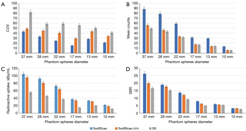
Methods: 99mTc-MDP SPECT/CT scans were acquired from the National Electrical Manufacturers Association (NEMA) phantom with a sphere-to-background ratio of 10:1, and 69 patients were enrolled in this retrospective study. The patients who showed abnormal uptake of focal 99mTc-MDP on whole-body bone planar scans, underwent local SPECT/CT scans. The image comparison and analysis were performed in three acquisition modes: SwiftScan; SwiftScan-50% (the SwiftScan mode with a 50% reduction in acquisition time); and step-and-shoot (SS). The image mean count, coefficient of variation (COV), radioactive concentration, and signal-to-background ratio (SBR) were measured for each phantom image. Visual assessment (using a 5-point Likert scale), semi-quantitative analysis [to determine the COV, mean of the standard uptake value (SUVmean), maximum of the standard uptake value (SUVmax), and SBR], and receiver operating characteristics (ROC) curve analysis were performed for clinical assessment.
Results: The SwiftScan mode obtained better quality images than the SwiftScan-50% and SS modes, and it also had better sphere clarity and lower background COVs (P<0.05). The mean counts of the SwiftScan, and the corresponding radioactive concentration and SBR were significantly higher than those of the SwiftScan-50% and SS (P<0.05), except in relation to the 13-10 mm spheres. A total of 101 abnormal 99mTc-MDP uptake skeletal lesions were included in the clinical study. The SwiftScan had significantly higher visual scores than both the SwiftScan-50% and SS (P<0.001). Additionally, there were significantly higher visual scores in the SwiftScan-50% than SS (P<0.05). The SwiftScan also had a lower COV, indicating reduced image noise. The SUVmax, SUVmean, and SBR for the skeletal lesions were significantly higher in the SwiftScan than the other two modes (P<0.05). The optimal diagnostic cut-off values of the SUVmax for identifying malignant skeletal lesions in the SwiftScan, SwiftScan-50%, and SS were 17.95, 14.75, and 9.94, respectively, but there was no significant difference among the three modes in terms of their differential diagnostic performance (all P>0.05).
Conclusions: The SwiftScan showed significant improvements in image quality and the semi-quantitative analysis of bone SPECT imaging. It reduced the scanning time by 50% without compromising the image quality or diagnostic performance. These findings provide valuable insights that may inform the clinical application of the SwiftScan, enhancing qualitative and quantitative diagnosis in bone SPECT imaging.
Keywords: SwiftScan; bone; image quality; semi-quantification; single-photon emission computed tomography/computed tomography (SPECT/CT).
2024 AME Publishing Company. All rights reserved.
Conflict of interest statement
Conflicts of Interest: All authors have completed the ICMJE uniform disclosure form (available at https://qims.amegroups.com/article/view/10.21037/qims-24-627/coif). The authors have no conflicts of interest to declare.
Figures


Similar articles
-
Verification of the effect of acquisition time for SwiftScan on quantitative bone single-photon emission computed tomography using an anthropomorphic phantom.EJNMMI Phys. 2022 Jul 30;9(1):48. doi: 10.1186/s40658-022-00477-9. EJNMMI Phys. 2022. PMID: 35907090 Free PMC article.
-
The SwiftScan step-and-shoot continuous mode improves SPECT scanning efficiency: a preliminary phantom and clinical test.EJNMMI Phys. 2025 Jan 2;12(1):1. doi: 10.1186/s40658-024-00709-0. EJNMMI Phys. 2025. PMID: 39745654 Free PMC article. Review.
-
Improving the image quality of short-time bone SPECT using cadmium-zinc-telluride detectors with SwiftScan.Asia Ocean J Nucl Med Biol. 2025;13(1):87-93. doi: 10.22038/aojnmb.2024.76919.1543. Asia Ocean J Nucl Med Biol. 2025. PMID: 39744056 Free PMC article.
-
The usefulness of SwiftScan technology for bone scintigraphy using a novel anthropomorphic phantom.Sci Rep. 2021 Jan 29;11(1):2644. doi: 10.1038/s41598-021-82082-x. Sci Rep. 2021. PMID: 33514818 Free PMC article.
-
Comparison of the diagnostic value of 18F-NaF PET/CT and 99mTc-MDP SPECT for bone metastases: a systematic review and meta-analysis.Transl Cancer Res. 2023 Nov 30;12(11):3166-3178. doi: 10.21037/tcr-23-817. Epub 2023 Nov 17. Transl Cancer Res. 2023. PMID: 38130318 Free PMC article. Review.
References
-
- Van den Wyngaert T, Strobel K, Kampen WU, Kuwert T, van der Bruggen W, Mohan HK, Gnanasegaran G, Delgado-Bolton R, Weber WA, Beheshti M, Langsteger W, Giammarile F, Mottaghy FM, Paycha F, Bone EANM, Joint Committee and the Oncology Committee . The EANM practice guidelines for bone scintigraphy. Eur J Nucl Med Mol Imaging 2016;43:1723-38. 10.1007/s00259-016-3415-4 - DOI - PMC - PubMed
-
- Gherghe M, Mutuleanu MD, Stanciu AE, Irimescu I, Lazar AM, Toma RV, Trifanescu OG, Anghel RM. Quantitative Assessment of Treatment Response in Metastatic Breast Cancer Patients by SPECT-CT Bone Imaging-Getting Closer to PET-CT. Cancers (Basel) 2023;15:696. 10.3390/cancers15030696 - DOI - PMC - PubMed
LinkOut - more resources
Full Text Sources