

Prenatal diagnosis of achondroplasia and hypochondroplasia using three-dimensional computed tomography: a case series at a single institution
- PMID: 39698715
- PMCID: PMC11651972
- DOI: 10.21037/qims-24-682
Prenatal diagnosis of achondroplasia and hypochondroplasia using three-dimensional computed tomography: a case series at a single institution
Abstract
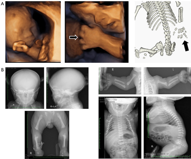
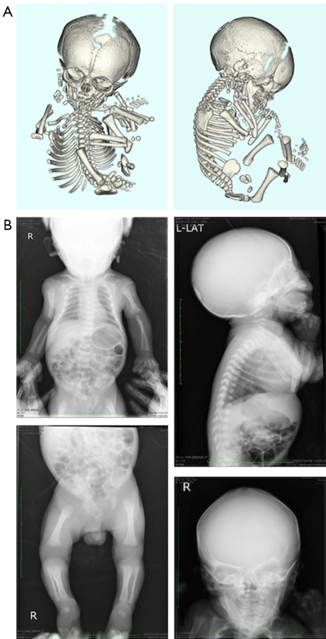
Background: Fetal skeletal dysplasia (FSD) is a group of systemic bone and cartilage disorders that develop prenatally and can be detected using fetal ultrasonography. However, it is unsuitable for skeletal analysis because it is reflected by supersonic waves in the bone cortex. Three-dimensional computed tomography (3D-CT) is a suitable alternative and has improved the differential diagnosis of FSD during pregnancy. Achondroplasia (ACH) and hypochondroplasia (HCH) are the most frequent diseases in the FSD group. This study aimed to determine the efficacy of 3D-CT in the prenatal diagnosis of ACH/HCH.
Case description: Patients were selected for the study non-consecutively. Pregnant women who met the following selection criteria were included: (I) pregnancy at age ≥20 years at the time of consent; (II) fetal ultrasonography showing a short femur (below the 10th percentile of fetal measurements) or an image of a long bone curvature or deformity; and (III) written informed consent to undergo 3D-CT. On suspicion of FSD based on ultrasonography, a 3D-CT scan was performed prenatally and an X-ray postnatally to obtain detailed skeletal information and to verify the shapes of all bones, respectively. A genetic examination was performed to confirm the diagnosis after obtaining informed consent from the parents. There were seven cases of prenatally diagnosed ACH/HCH. Genetic testing was performed in six infants postnatally, and a mutation in the fibroblast growth factor receptor 3 (FGFR3) gene was detected [c. 1138G>A (p. gly380Arg)]. In one case, the patient was diagnosed with ACH and Down syndrome by genetic and chromosomal testing (G-band: 47, XY, +21).
Conclusions: 3D-CT is a valuable and efficient tool for diagnosing ACH/HCH. Accurate prenatal diagnosis by gene analysis is crucial for a definitive diagnosis in infants.
Keywords: Achondroplasia (ACH); case series; fetal diagnosis; fetal skeletal dysplasia (FSD); three-dimensional computed tomography (3D-CT).
2024 AME Publishing Company. All rights reserved.
Conflict of interest statement
Conflicts of Interest: All authors have completed the ICMJE uniform disclosure form (available at https://qims.amegroups.com/article/view/10.21037/qims-24-682/coif). The authors have no conflicts of interest to declare.
Figures

Similar articles
-
Prenatal diagnosis of fetal skeletal dysplasia using 3-dimensional computed tomography: a prospective study.BMC Musculoskelet Disord. 2020 Oct 8;21(1):662. doi: 10.1186/s12891-020-03663-x. BMC Musculoskelet Disord. 2020. PMID: 33032557 Free PMC article.
-
Detection of a de novo Y278C mutation in FGFR3 in a pregnancy with severe fetal hypochondroplasia: prenatal diagnosis and literature review.Taiwan J Obstet Gynecol. 2013 Dec;52(4):580-5. doi: 10.1016/j.tjog.2013.10.023. Taiwan J Obstet Gynecol. 2013. PMID: 24411048 Review.
-
Low bone mineral density in achondroplasia and hypochondroplasia.Pediatr Int. 2016 Aug;58(8):705-8. doi: 10.1111/ped.12890. Epub 2016 Apr 5. Pediatr Int. 2016. PMID: 26716907
-
p.Ser348Cys mutation in FGFR3 gene leads to "Mild ACH /Severe HCH" phenotype.Eur J Med Genet. 2020 Feb;63(2):103659. doi: 10.1016/j.ejmg.2019.04.016. Epub 2019 Apr 30. Eur J Med Genet. 2020. PMID: 31048079
-
Earlier detection of hypochondroplasia: A large single-center UK case series and systematic review.Am J Med Genet A. 2021 Jan;185(1):73-82. doi: 10.1002/ajmg.a.61912. Epub 2020 Oct 14. Am J Med Genet A. 2021. PMID: 33051983
References
-
- Schumacher R, Seaver LH, Spranger J. Fetal Radiology A Diagnostic Atlas. Springer-verlag, Berlin, 2004.
Publication types
LinkOut - more resources
Full Text Sources
Miscellaneous