

doi: 10.21037/qims-24-1101.
Epub 2024 Nov 29.
Ectopic pituitary adenoma in sphenoid sinus: a case presentation
Affiliations
- PMID: 39698731
- PMCID: PMC11652051
- DOI: 10.21037/qims-24-1101
Item in Clipboard
Ectopic pituitary adenoma in sphenoid sinus: a case presentation
Quant Imaging Med Surg.
.
No abstract available
Conflict of interest statement
Conflicts of Interest: Both authors have completed the ICMJE uniform disclosure form (available at https://qims.amegroups.com/article/view/10.21037/qims-24-1101/coif). The authors have no conflicts of interest to declare.
Figures
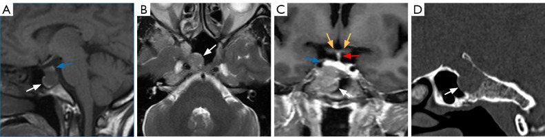
Preoperative MRI and CT images of the tumor. (A) Sagittal position on T1WI, an irregular low-signal shadow (white arrow) could be observed in the sphenoid sinus. Blue arrow: pituitary gland. (B) Transverse position on T2WI, an irregular isointense shadow (white arrow) can be seen; (C) coronal position of T1 contrast enhancement. An irregular soft tissue mass (white arrow) was observed in the sphenoid sinus with mild enhancement (white arrow), with unclear boundary of the right sellar bottom, and pituitary gland located at the superior parts of the lesion. Yellow arrows: optic chiasm; blue arrow: pituitary gland; red arrow: infundibulum. (D) CT bone window scan showed the soft tissue shadow (white arrow) and bone destruction. MRI, magnetic resonance imaging; CT, computed tomography; T1WI, T1-weighted imaging; T2WI, T2-weighted imaging.
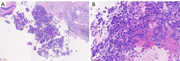
HE stain of the EPAs. (A) HE ×100, the tumor cells consisted of uniform, small, round cells arranged in nests under microscopy. (B) HE ×400, the tumor cells are separated by a network of slender blood vessels, the cells are rich in the cytoplasm and arranged in a nested, glandular pattern, and the nuclei have fine chromatin. HE, hematoxylin and eosin; EPAs, ectopic pituitary adenomas.
Similar articles
-
Three cases of ectopic sphenoid sinus pituitary adenoma.Folia Neuropathol. 2017;55(1):60-66. doi: 10.5114/fn.2017.66714. Folia Neuropathol. 2017. PMID: 28430293
-
[A case of ectopic pituitary adenoma occurring in the sphenoid sinus].No Shinkei Geka. 2011 Jun;39(6):601-5. No Shinkei Geka. 2011. PMID: 21628740 Japanese.
-
[Ectopic pituitary tumor of sphenoid sinus: a case report].Lin Chuang Er Bi Yan Hou Tou Jing Wai Ke Za Zhi. 2024 Jun;38(6):553-555. doi: 10.13201/j.issn.2096-7993.2024.06.018. Lin Chuang Er Bi Yan Hou Tou Jing Wai Ke Za Zhi. 2024. PMID: 38858124 Free PMC article. Chinese.
-
Ectopic acromegaly due to a GH-secreting pituitary adenoma in the sphenoid sinus: a case report and review of the literature.BMC Res Notes. 2013 Oct 12;6:411. doi: 10.1186/1756-0500-6-411. BMC Res Notes. 2013. PMID: 24119925 Free PMC article. Review.
-
Ectopic ACTH-secreting pituitary adenomas within the sphenoid sinus.Endocrine. 2014 Dec;47(3):717-24. doi: 10.1007/s12020-014-0313-z. Epub 2014 Jun 14. Endocrine. 2014. PMID: 24927792 Review.
References
-
- Campana C, Nista F, Castelletti L, Caputo M, Lavezzi E, Marzullo P, Ferrero A, Gaggero G, Canevari FR, Rossi DC, Zona G, Lania A, Ferone D, Gatto F. Clinical and radiological presentation of parasellar ectopic pituitary adenomas: case series and systematic review of the literature. J Endocrinol Invest 2022;45:1465-81. 10.1007/s40618-022-01758-x - DOI - PubMed
LinkOut - more resources
Full Text Sources