. 2025 Jan 24;53(3):gkae1248.
doi: 10.1093/nar/gkae1248.

RNA elements required for the high efficiency of West Nile virus-induced ribosomal frameshifting

Affiliations

RNA elements required for the high efficiency of West Nile virus-induced ribosomal frameshifting

Nikolay A Aleksashin et al. Nucleic Acids Res. .

Abstract

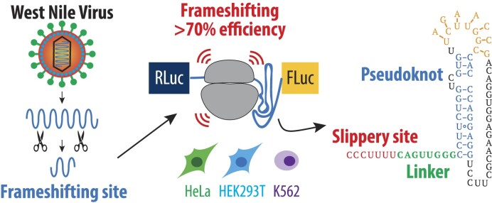
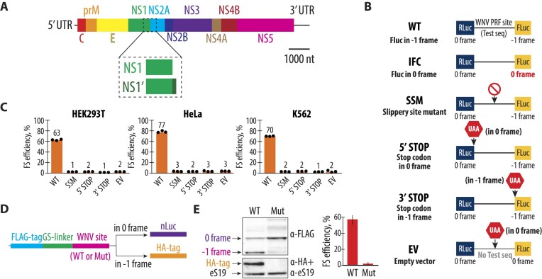
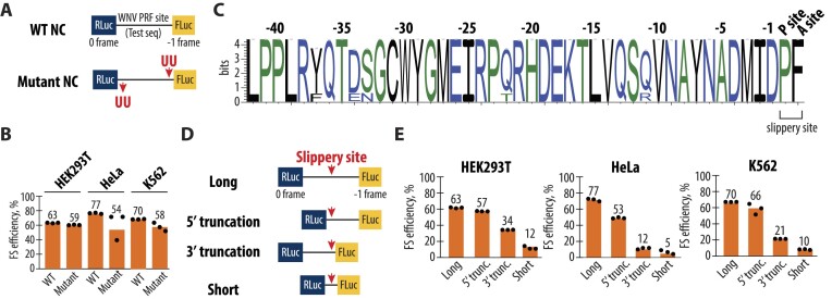
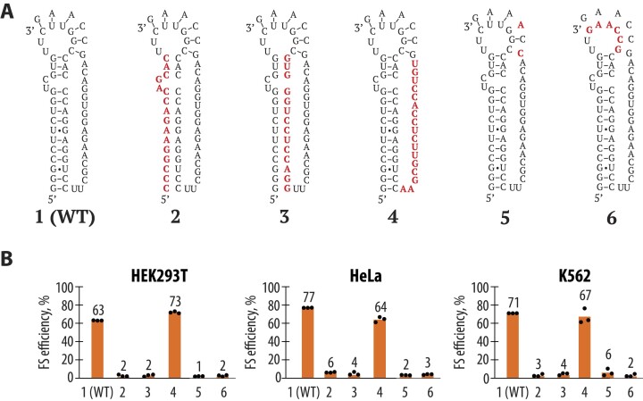
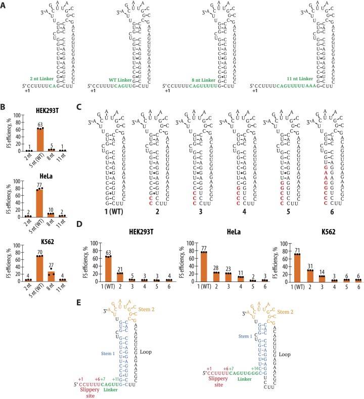
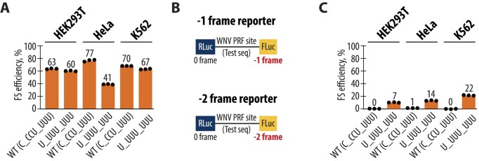
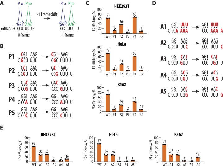
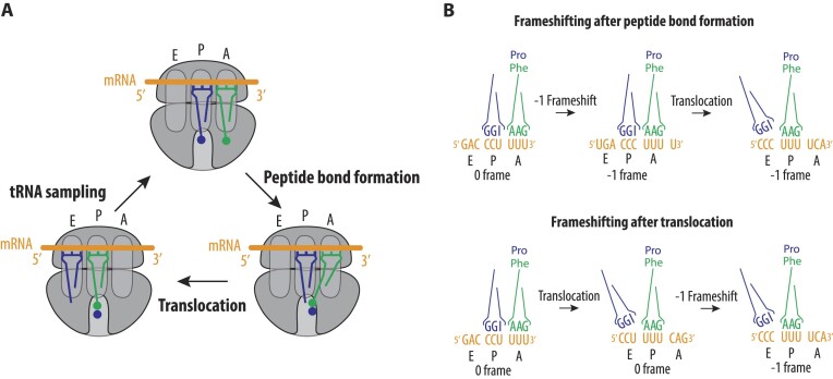
West Nile virus (WNV) requires programmed -1 ribosomal frameshifting for translation of the viral genome. The efficiency of WNV frameshifting is among the highest known. However, it remains unclear why WNV exhibits such a high frameshifting efficiency. Here, we employed dual-luciferase reporter assays in multiple human cell lines to probe the RNA requirements for highly efficient frameshifting by the WNV genome. We find that both the sequence and structure of a predicted RNA pseudoknot downstream of the slippery sequence-the codons in the genome on which frameshifting occurs-are required for efficient frameshifting. We also show that multiple proposed RNA secondary structures downstream of the slippery sequence are inconsistent with efficient frameshifting. We also find that the base of the pseudoknot structure likely is unfolded prior to frameshifting. Finally, we show that many mutations in the WNV slippery sequence allow efficient frameshifting, but often result in aberrant shifting into other reading frames. Mutations in the slippery sequence also support a model in which frameshifting occurs concurrent with or after ribosome translocation. These results provide a comprehensive analysis of the molecular determinants of WNV-programmed ribosomal frameshifting and provide a foundation for the development of new antiviral strategies targeting viral gene expression.


Figures

Graphical Abstract
Graphical Abstract
Figure 1.
Figure 1.
Programmed −1 ribosomal frameshifting at the NS1 and NS2A regions of the WNV genome. (A) Schematic representation of the WNV genome. The genome consists of a single positive-sense RNA that encodes a polyprotein, which is co- and post-translationally processed into three structural proteins (C, prM and E) and seven nonstructural proteins (NS1, NS2A, NS2B, NS3, NS4A, NS4B and NS5). The NS1 and NS2A junction includes the programmed −1 ribosomal frameshifting site, which leads to the production of both the full-length NS1 and the extended NS1′ protein. (B) Diagram of the dual-luciferase reporter constructs used to measure frameshifting efficiency. The wild-type (WT) reporter contains Renilla luciferase (RLuc) in the reference reading frame (0 frame) and firefly luciferase (FLuc) in the −1 reading frame, separated by the WNV PRF site. The IFC construct controls for no frameshifting by placing FLuc in the 0 frame. The mutations in the slippery site (SSM) are designed to abolish frameshifting. The 5′ STOP and 3′ STOP constructs introduce stop codons in either the 0 or −1 frames before or after the testing sequence, respectively, while the empty vector (EV) contains no test sequence. See the ‘Materials and methods’ section for more on the use of the IFC construct for normalizing experiments. (C) The −1 frameshifting efficiencies of the WT and mutant constructs in HEK293T, HeLa and K562 cell lines. Frameshifting efficiency is shown as a percentage of the ratio between normalized FLuc/RLuc luminescence in the −1 frame and the sum of normalized Fluc/Rluc luminescence in −1 and 0 frames. Bars represent the means of three independent experiments, with each dot corresponding to the individual experimental data points. (D) Schematic of the reporter construct used in the in vitro translation system based on human extracts (39). The reporters include either WT or mutant (Mut) WNV frameshifting sites. While the translation products always carry a 3x FLAG tag followed by GS linker on their N-terminus, only −1 frameshifting product polypeptides have an HA tag on their C-terminus. Ribosomes that continue translation in the 0 frame synthesize nanoluciferase (nLuc). (E) Western blot analysis of FLAG and HA tags in WT and mutant constructs after in vitro translation reactions. The WT construct shows both FLAG- and HA-tagged proteins, indicating efficient frameshifting. The Mut construct abolishes frameshifting, as indicated by the absence of the HA-tagged product, while increasing nLuc protein levels as detected by the α-FLAG band. Quantification of frameshifting (FS) efficiency is shown on the right.
Figure 2.
Figure 2.
Importance of the nascent peptide and mRNA length for the ribosome frameshifting. (A) Schematic representation of the WT and mutant 2-nt register-shifted reporter constructs. In the WT construct, the FLuc gene is positioned in the −1 reading frame relative to the RLuc gene, separated by the WNV PRF site. In the mutant register-shifted construct, two U residues deleted at the beginning of the testing sequence and inserted back upstream of the frameshifting site change the nascent peptide sequence while having a minimal effect on the mRNA sequence. Five additional nucleotide changes that remove potential stop codons in the 2-nt register-shifted sequence are shown in Supplementary Figure S2. (B) Frameshifting efficiencies of the WT and mutant constructs measured in HEK293T, HeLa and K562 cell lines. Frameshifting efficiency is shown as a percentage of the ratio between normalized FLuc/RLuc luminescence in the −1 frame and the sum of normalized Fluc/Rluc luminescence in the −1 and 0 frames. Bars represent the means of three independent experiments, with each dot corresponding to the individual experimental data points. (C) Sequence logo information content in bits of the 0-frame nascent peptide multiple sequence alignment immediately upstream from the WNV PRF site, for all complete WNV genomes deposited in the NCBI sequence database. The height of each position represents the relative information content of each position in bits and the x-axis displays the relative position of the amino acid in the multiple sequence alignment. (D) Schematic of truncation constructs used to assess the effect of sequences surrounding the slippery sequence site on frameshifting efficiency. The ‘Long’ construct includes the full-length PRF sequence (258 nt), while the ‘5′ truncation’ (135 nt), ‘3′ truncation’ (204 nt) and ‘Short’ (81 nt) constructs progressively reduce the sequence length around the slippery site. (E) Frameshifting efficiencies for the truncation constructs in HEK293T, HeLa and K562 cells. Frameshifting efficiency is shown as a percentage of the ratio between normalized FLuc/RLuc luminescence in the −1 frame and the sum of normalized Fluc/Rluc luminescence in the −1 and 0 frames. Bars represent the means of three independent experiments, with each dot corresponding to the individual experimental data points.
Figure 3.
Figure 3.
Mutational probing of the pseudoknot structure in WNV −1 frameshifting. (A) Schematic representation of the mutations designed to disrupt the predicted pseudoknot structure. Mutations are shown in bold and were targeted to various regions of the pseudoknot, including the stem and loop regions. (B) Results of the frameshifting efficiency assays for the WT and mutant pseudoknot constructs in HEK293T, HeLa and K562 cells. Frameshifting efficiency is shown as a percentage of the ratio between normalized FLuc/RLuc luminescence in the −1 frame and the sum of normalized Fluc/Rluc luminescence in the −1 and 0 frames. Bars represent the means of three independent experiments, with each dot corresponding to the individual experimental data points.
Figure 4.
Figure 4.
Analysis of linker length and pseudoknot structure of the WNV frameshifting site. (A) Schematic representation of the mutations altering the length of the linker region between the slippery site and the pseudoknot. (B) Frameshifting efficiency of the WNV sequence with different linker lengths in HEK293T, HeLa and K562 cells. Frameshifting efficiency is shown as a percentage of the ratio between normalized FLuc/RLuc luminescence in the −1 frame and the sum of normalized Fluc/Rluc luminescence in the −1 and 0 frames. Bars represent the means of three independent experiments, with each dot corresponding to the individual experimental data points. (C) Representation of the WT pseudoknot structure and mutations that disrupt predicted base pairs at the base of the pseudoknot stem (mutations highlighted in bold). Constructs are numbered 1 (i.e. WT) through 6. (D) Frameshifting efficiency of the mutant sequences with disrupted base pairs compared to WT, in HEK293T, HeLa and K562 cells. Frameshifting efficiency is shown as a percentage of the ratio between normalized FLuc/RLuc luminescence in the −1 frame and the sum of normalized Fluc/Rluc luminescence in the −1 and 0 frames. Bars represent the means of three independent experiments, with each dot corresponding to the individual experimental data points. (E) Proposed structure of the WNV frameshifting site based on the reporter assays (right), compared to the previously predicted structure (left).
Figure 5.
Figure 5.
Effect of slippery site sequences on frameshifting efficiency and register in the WNV PRF site. (A) Frameshifting efficiencies of the WT (C_CCU_UUU) and mutant (U_UUU_UUU) frameshifting constructs in HEK293T, HeLa and K562 cell lines. Frameshifting efficiency is shown as a percentage of the ratio between normalized FLuc/RLuc luminescence in the −1 frame and the sum of normalized Fluc/Rluc luminescence in the −1 and 0 frames. (B) Schematic representation of the dual-luciferase reporters designed to measure −1 and −2 ribosomal frameshifting. In the −1 frame reporter, FLuc is positioned in the −1 frame, whereas in the −2 frame reporter, FLuc is positioned in the −2 frame relative Rluc in the 0 frame. (C) Efficiency of −2 ribosomal frameshifting in HEK293T, HeLa and K562 cells for the WT and mutant constructs. The WT sequence shows no detectable −2 frameshifting, while the mutant (U_UUU_UUU) construct induces low levels of −2 frameshifting, particularly in K562 cells. Frameshifting efficiency is shown as a percentage of the ratio between normalized FLuc/RLuc luminescence in the −2 frame and the sum of normalized Fluc/Rluc luminescence in the −2 and 0 frames. In panels (A) and (C), bars represent the means of three independent experiments, with each dot corresponding to the individual experimental data points.
Figure 6.
Figure 6.
Mutational analysis of codon–anticodon interactions during WNV frameshifting. (A) Overview of codon–anticodon interactions before and after the −1 frameshift. P-site tRNA Pro (on the left) and A-site tRNA Phe (on the right) in the example of the WT WNV slippery site are shown interacting with their codons. (B) Codon–anticodon interactions for constructs with mutations in the P-site codon before and after frameshifting. The mRNA mutations and corresponding changes in the anticodon sequences of tRNAs are shown in bold. (C) Frameshifting efficiency for the P-site mutant constructs in HEK293T, HeLa and K562 cells. Frameshifting efficiency is shown as a percentage of the ratio between normalized FLuc/RLuc luminescence in the −1 frame and the sum of normalized Fluc/Rluc luminescence in the −1 and 0 frames. (D) Codon–anticodon interactions for constructs with mutations in the A-site codon before and after frameshifting. The mRNA mutations and corresponding changes in the anticodon sequences of tRNAs are shown in bold. (E) Frameshifting efficiency for the A-site mutant constructs in HEK293T, HeLa and K562 cells. Frameshifting efficiency is shown as a percentage of the ratio between normalized FLuc/RLuc luminescence in the −1 frame and the sum of normalized Fluc/Rluc luminescence in the −1 and 0 frames. In panels (C) and (E), the bars represent the means of three independent experiments, with each dot corresponding to the individual experimental data points.
Figure 7.
Figure 7.
Model of WNV −1 frameshifting. (A) The regular elongation cycle consists of tRNA sampling, peptide bond formation and translocation, with ribosomes spontaneously changing reading frames at a very low rate (∼0.01%) (85–87). (B) Possible models of frameshifting induced by the WNV PRF site: (i) Frameshifting may happen after peptide bond formation but before the ribosome undergoes translocation. (ii) Frameshifting may alternatively occur after translocation and E-site tRNA dissociation, when only one tRNA occupies the P site. This would establish the −1 frame for the next mRNA decoding event in the A site. Not shown, frameshifting could also take place simultaneously with the translocation event, resulting in the ribosome shifting by one nucleotide to the −1 frame, with the tRNAs positioned in the E site and P site.

Update of

Similar articles

Cited by

References

    1. Atkins J.F., Loughran G., Bhatt P.R., Firth A.E., Baranov P.V.. Ribosomal frameshifting and transcriptional slippage: from genetic steganography and cryptography to adventitious use. Nucleic Acids Res. 2016; 44:7007–7078. - PMC - PubMed
    1. Firth A.E., Brierley I.. Non-canonical translation in RNA viruses. J. Gen. Virol. 2012; 93:1385–1409. - PMC - PubMed
    1. Hayes E.B., Komar N., Nasci R.S., Montgomery S.P., O’Leary D.R., Campbell G.L.. Epidemiology and transmission dynamics of West Nile virus disease. Emerg. Infect. Dis. 2005; 11:1167–1173. - PMC - PubMed
    1. Martín-Acebes M.A., Saiz J.-C.. West Nile virus: a re-emerging pathogen revisited. World J. Virol. 2012; 1:51. - PMC - PubMed
    1. Smithburn K.C., Hughes T.P., Burke A.W., Paul J.H.. A neurotropic virus isolated from the blood of a native of Uganda. Am. J. Trop. Med. 1940; s1-20:471–492.