. 2025 Jan 11;53(2):gkae1198.
doi: 10.1093/nar/gkae1198.

Structural plasticity of the coiled-coil interactions in human SFPQ

Affiliations

Structural plasticity of the coiled-coil interactions in human SFPQ

Heidar J Koning et al. Nucleic Acids Res. .

Abstract

The proteins SFPQ (splicing Factor Proline/Glutamine rich) and NONO (non-POU domain-containing octamer-binding protein) are mammalian members of the Drosophila Behaviour/Human Splicing (DBHS) protein family, which share 76% sequence identity in their conserved 320 amino acid DBHS domain. SFPQ and NONO are involved in all steps of post-transcriptional regulation and are primarily located in mammalian paraspeckles: liquid phase-separated, ribonucleoprotein sub-nuclear bodies templated by NEAT1 long non-coding RNA. A combination of structured and low-complexity regions provide polyvalent interaction interfaces that facilitate homo- and heterodimerisation, polymerisation, interactions with oligonucleotides, mRNA, long non-coding RNA, and liquid phase-separation, all of which have been implicated in cellular homeostasis and neurological diseases including neuroblastoma. The strength and competition of these interaction modes define the ability of DBHS proteins to dissociate from paraspeckles to fulfil functional roles throughout the nucleus or the cytoplasm. In this study, we define and dissect the coiled-coil interactions which promote the polymerisation of DBHS proteins, using a crystal structure of an SFPQ/NONO heterodimer which reveals a flexible coiled-coil interaction interface which differs from previous studies. We support this through extensive solution small-angle X-ray scattering experiments using a panel of SFPQ/NONO heterodimer variants which are capable of tetramerisation to varying extents. The QM mutant displayed a negligible amount of tetramerisation (quadruple loss of function coiled-coil mutant L535A/L539A/L546A/M549A), the Charged Single Alpha Helix (ΔCSAH) variant displayed a dimer-tetramer equilibrium interaction, and the disulfide-forming variant displayed constitutive tetramerisation (R542C which mimics the pathological Drosophila nonAdiss allele). We demonstrate that newly characterised coiled-coil interfaces play a role in the polymerisation of DBHS proteins in addition to the previously described canonical coiled-coil interface. The detail of these interactions provides insight into a process critical for the assembly of paraspeckles as well as the behaviour of SFPQ as a transcription factor, and general multipurpose auxiliary protein with functions essential to mammalian life. Our understanding of the coiled coil behaviour of SFPQ also enhances the explanatory power of mutations (often disease-associated) observed in the DBHS family, potentially allowing for the development of future medical options such as targeted gene therapy.


Figures

Graphical Abstract
Graphical Abstract
Figure 1.
Figure 1.
Crystal structure of an SFPQ/NONO heterodimer. (A) Novel crystal structure of an SFPQ/NONO heterodimer colored yellow (RRM1), dark blue (RRM2), orange (NOPS), red (coiled-coil or CC) and gold (CSAH) of SFPQ in cartoon representation. NONO is shown in surface representation. The construct boundaries used in this study are drawn and include residues 214–598 of SFPQ and 53–312 of NONO with the colours matching the crystal structure. (B) The aligned amino acid sequences of the coiled–coil domain in NONO-1 from C. elegans, SFPQ, PSPC1, and NONO from H. sapiens. These are colored by amino acid similarity (darker colours indicate amino acid agreement). The Alphafold predicted α-helical regions are indicated below with the canonical coiled–coil interface region in red and the CSAH region in gold. The region beyond the coiled–coil interaction motif in NONO-1 is not predicted to be alpha helical and lacks homology to the CSAH region in the human paralogs.
Figure 2.
Figure 2.
Crystal structure of an SFPQ/NONO tetramer. (A) Superposition of the two heterodimers of SFPQ/NONO in the asymmetric unit reveals the domain movement of RRM1 and the CC domain of SFPQ. Chain A (SFPQ) in yellow, Chain B (NONO) in pale pink, Chain C (SFPQ) in cyan and Chain D (NONO) in grey. (B) Tetramer interface formed in the asymmetric unit of the crystal, Chain A (SFPQ) in yellow, Chain B (NONO) in pale pink, Chain C (SFPQ) in cyan, and Chain D (NONO) in grey. (C) Close-up view of the CC interaction mediated dimer-dimer interface. (D) Comparison with the CC interaction interface with that in the SFPQ polymer (4WIJ) using the same viewpoint as in the lower panel of (B). The coiled–coil interaction motif (residues 528–555) of the SFPQ/NONO tetramer (black) and that in Chain A of the SFPQ homodimer tetramer structure (grey) (4WIJ) are superposed. The neighboring symmetry-related Chain B (y + 1/2, -x + 1/2, z + 1/4) of the SFPQ homodimer structure (4WIJ) is shown while NONO is omitted in this figure for simplicity.
Figure 3.
Figure 3.
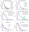
Solution studies on protein variants of SFPQ/NONO (A) Boundaries of all the SFPQ constructs used in SAXS experiments and attached descriptions of their observed behaviour in solution. (B) Log(I) versus Log(q) plot for SFPQ276-565/NONO53-312 (ΔCSAH) alongside a residual plot describing the CRYSOL fit of a truncated 6WMZ model to the data. (C) Log(I) versus Log(q) plot for SFPQ276-598/NONO53-312(QM) alongside a residual plot describing the CRYSOL fit of a 6WMZ model to the data. Systematic wavelike deviations in the residuals plot are likely caused by movement of the coiled–coil domain in solution (D) Log(I) versus Log(q) plot for SFPQ276-598(R542C)/NONO260. (E) Log (I) versus Log(q) plot for SFPQ214-598(R542C)/NONO53-312. Data were captured using static-SAXS and SEC-SAXS, respectively. (F) Guinier fits calculated in BioXTAS RAW with the associated Rg values for each construct in angstroms. The ΔCSAH residuals were flat across q.Rg 0.3119–1.2822 suggesting a globular particule. The QM residuals began to uptick at q.Rg > 1 and so the Guinier fitting range was adjusted to q.Rg 0.228–1.01 and was linear suggesting a partly rodlike particle. The SEC-SAXS data (R542C-DBD) showed flat residuals over the q.Rg range 0.201–1.284, suggesting a globular particle. Given this q.Rg max was set as 1.3 for the R542C alone fit and the first five data points excluded from the fit (35). However, the residuals still showed a characteristic smile pattern suggesting aggregation.
Figure 4.
Figure 4.
Dimers and a constitutive tetramer of SFPQ/NONO (A) CHROMIXS SEC-SAXS chromatography trace for SFPQ214-598(R542C)/NONO53-312. Scattering intensity is plotted in blue with the estimated radius of gyration plotted in red. (B) P(r) functions for the constructs considered amenable to 3D reconstruction plotted against theoretical distributions to assess homogeneity and structure. (C) Kratky plot of 3D modelling candidates to assess foldedness and globularity. (D/E) DAMAVER (grey) and DAMFILT (cyan) models of the QM and ΔCSAH constructs. Black crystal structure of the 6WMZ dimer with a full-length coiled–coil domain and truncated version superposed over the bead models. (F) GASBOR model generated from the SFPQ214-598(R542C)/NONO53-312 dataset demonstrates that the data roughly correspond to the shape of a tetramer. (G) pLDDT scores for different models of SFPQ214-598 produced in CollabFold. This can be considered a relatively strong indicator that the first 65 residues of this construct are likely disordered (scores below or close to 0.5). (H/I) CORAL fits to the data of SFPQ214-598(R542C)/NONO53-312 demonstrate that a variety of input structures demonstrate different fits to the data. The blue model is derived from the dimer in the asymmetric unit of 6WMZ, which has a relatively straight coiled–coil domain. The grey model is the 6WMZ tetramer fixed as a rigid body. The pink model uses the other dimer from the asymmetric unit of 6WMZ which has a bent coiled–coil domain. Modelling constraints reflected a disulphide bond between coiled–coil domains as well as adjacent residues kept at ∼15 angstroms to mimic a canonical antiparallel coiled–coil interaction. Normalised residual plot indicating systematic deviations of the fit from the data, most likely caused by domain flexibility.
Figure 5.
Figure 5.
Distance distribution analysis of SFPQ variants and SVD (A) P(r) distribution of the SFPQ276-565/NONO53-312 dataset, curves indicate a change in structure as a function of concentration that resembles tetramerisation (9). Theoretical distributions are plotted alongside the data as a reference. (B) P(r) distributions for the SFPQ276-598/NONO53-312 (QM) dataset. Data indicate some concentration-dependent changes. (C) P(r) distributions for SFPQ276-565/NONO53-312 (ΔCSAH) and SFPQ276-598/NONO53-312 (WT) at comparable concentrations, a smaller distribution for SFPQ276-565/NONO53-312 at a higher concentration corresponds to a reduction in tetramerisation. (D) P(r) distribution for a dataset of SFPQ276-598/NONO53-312 plotted against the theoretical distributions of the 6WMZ tetramer and a tetramer formed via the non-canonical 4WIK interface. Green highlights represent predicted distances present in the experimental data and the 4WIK interface but absent from the theoretical distribution of a canonical tetramer. (E) Singular values obtained by SVD in BioXTAS Raw for all scattering experiments. (F) SVD autocorrelation results from BioXTAS RAW. Every data point above an absolute value of ∼0.6 is an estimated unique scattering species in solution.
Figure 6.
Figure 6.
The contribution of a multiple flexible coiled–coil interfaces to nucleic acid binding (A) A cartoon indicating, relative flexibilities of DBHS dimers and DNA, and DBHS-DNA filaments assisted by local flexibility in the coiled–coil domain. (B) tandem recognition of nucleic acids sites by the canonical coiled-coil ‘molecular ruler’, tandem recognition of nucleic acids sites by the hypothesised CSAH ‘molecular ruler’ and a hybrid interaction schematic allowing bridging of DBHS-nucleic acid complexes as posited by the birds nest model of Jiang et al. (56).

Similar articles

Cited by

References

    1. Knott G.J., Bond C.S., Fox A.H.. The DBHS proteins SFPQ, NONO and PSPC1: a multipurpose molecular scaffold. Nucleic Acids Res. 2016; 44:3989–4004. - PMC - PubMed
    1. Fox A.H., Nakagawa S., Hirose T., Bond C.S.. Paraspeckles: where long noncoding RNA meets phase separation. Trends Biochem. Sci. 2018; 43:124–135. - PubMed
    1. Marshall A.C., Cummins J., Kobelke S., Zhu T., Widagdo J., Anggono V., Hyman A., Fox A.H., Bond C.S., Lee M.. Different low-complexity regions of SFPQ play distinct roles in the formation of biomolecular condensates. J. Mol. Biol. 2023; 435:168364. - PubMed
    1. Huang J., Ringuet M., Whitten A.E., Caria S., Lim Y.W., Badhan R., Anggono V., Lee M.. Structural basis of the zinc-induced cytoplasmic aggregation of the RNA-binding protein SFPQ. Nucleic Acids Res. 2020; 48:3356–3365. - PMC - PubMed
    1. Lim Y.W., James D., Huang J., Lee M.. The emerging role of the RNA-binding protein SFPQ in neuronal function and neurodegeneration. Int. J. Mol. Sci. 2020; 21:7151. - PMC - PubMed

MeSH terms

LinkOut - more resources